
|
Fig. 1
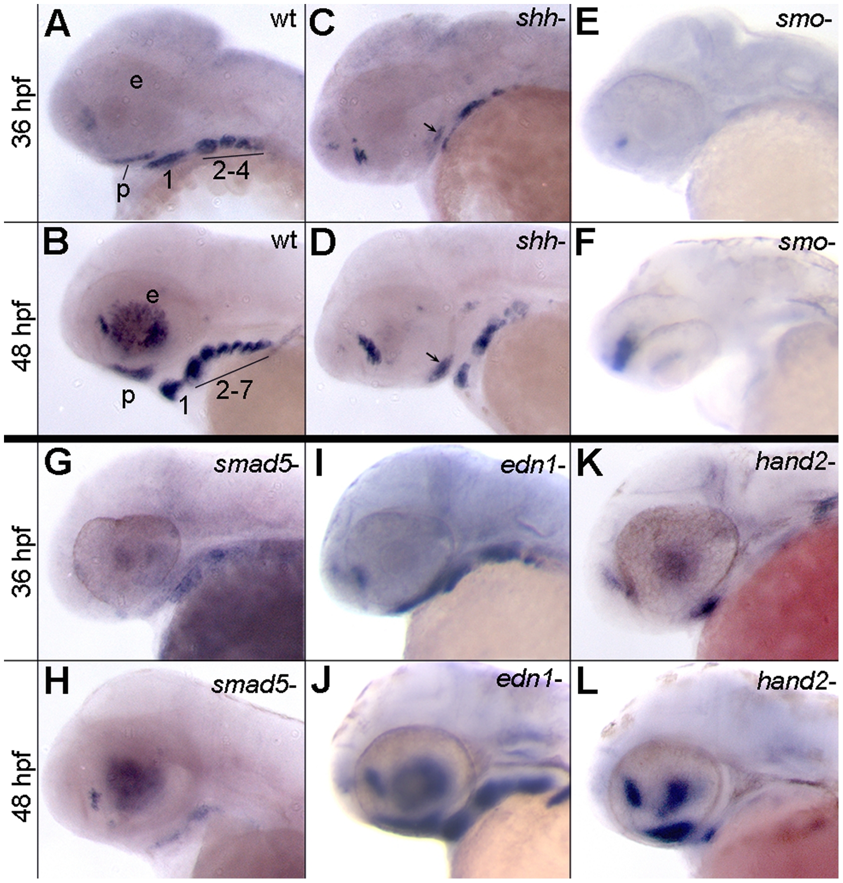
Expression of satb2 requires Hh and Bmp signaling.
(A–L) Lateral images of satb2 expression at 36 or 48 hpf. (A–D) Compared to wild-type embryos, in 36 and 48 hpf shha embryos, satb2 expression is reduced in the palatal precursors (arrows in C and D) and throughout the pharyngeal arches. (E,F) Nearly all neural crest cell expression of satb2 appears to be absent in smo embryos. The prominent expression in panel F, is in neural tissue. (G,H) Weak to no expression of satb2 was observed in smad5 mutant embryos at 36 and 48 hpf. (I,J) satb2 expression in edn1 mutants was similar wild-type embryos at both time points analyzed. (K,L) While expression of satb2 was absent throughout the posterior arches of hand2 mutants, the ventral first arch and palatal precursors maintained satb2 expression. p, palatal precursors; e, eye; The arches are numbered in A & C for clarity.