
|
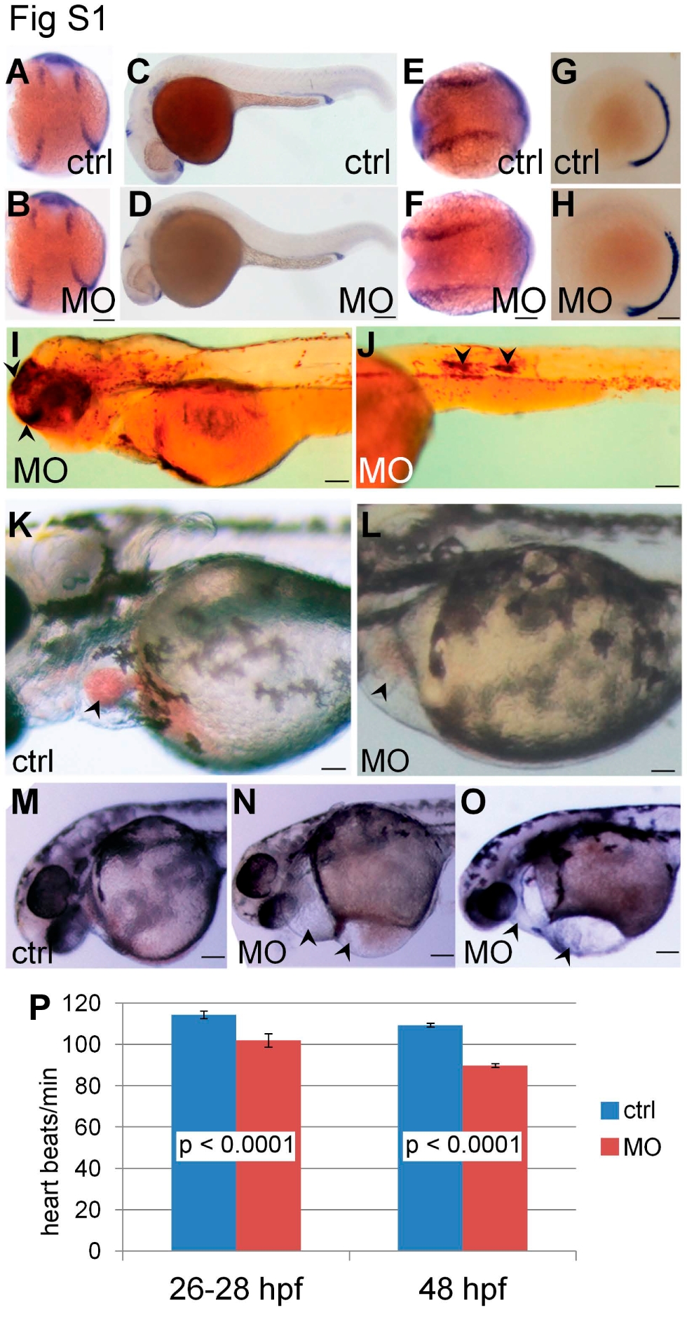
Fig. S1
Patterning and morphology in hoxd4a morphants. (A–H) Knockdown of hoxd4a does not perturb overall patterning of the mesoderm. (A–D) Expression of pax2.1 shows that intermediate mesoderm forms and matures normally in hoxd4a morphants at 13 hpf (B) and 26–28 hpf (D) vs controls (A,C). (E,F) nkx2.5 expression in the precardiac lateral plate mesoderm at 13 hpf is normal in control (E) and morphant embryos (F). (G,H) myod expression in paraxial mesoderm is normal in control (G) and morphant embryos (H). Images in C, D, G and H are lateral views with anterior to the left. A, B, E and F show dorsal views with anterior to the top (A,B) or left (E,F). (I to O) Lateral views of control and hoxd4a-MO-injected larvae at 72 hpf. (I,J) Staining of hemoglobin with o-dianisidine reveals areas of hemorrhage such as in the head (I, arrowheads) and trunk (J, arrowheads) in some hoxd4a morphants. (K,L) Control larvae (K) but not hoxd4a morphants (L) show abundant RBCs passing through the heart (arrowheads). (M–O) Unlike control larvae (M), hoxd4a morphants display pericardial edema and edema over the adjacent yolk (N,O, arrowheads). Scale bars equal 100 μm. (P) The heart rate in morphants at 26–28 and 48 hpf was mildly reduced, but in a statistically significant manner as determined by unpaired Student’s t test (p<0.0001). Error bars give standard deviation.