
|
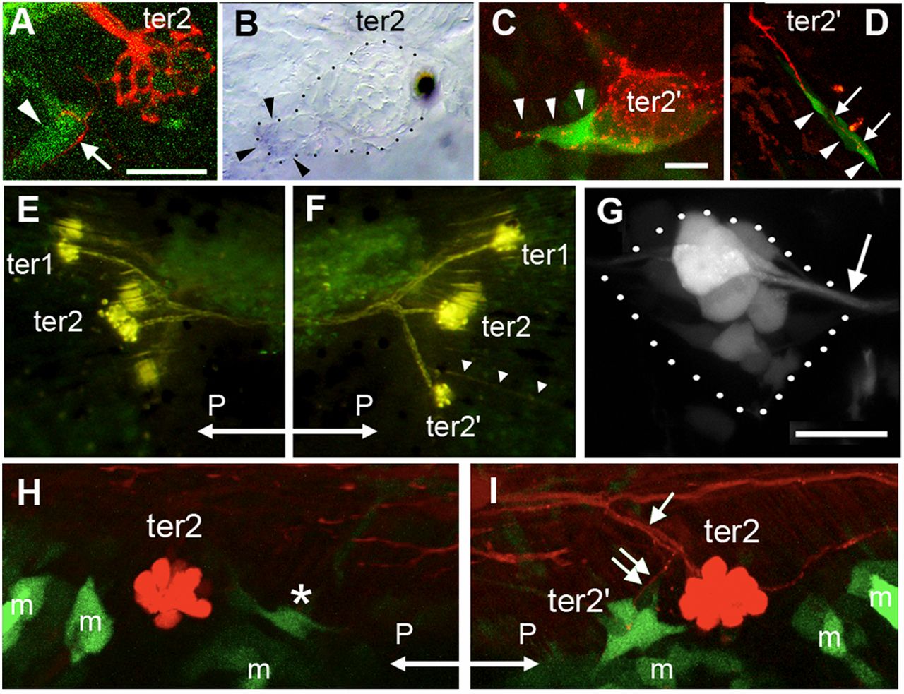
Fig. 4 Reporting Wnt and Rspo2 activity during budding. (A) Wnt activity (green) prefiguring ter2′, next to ter2, in a 9-dpf larva. Wnt activity (arrowhead) is adjacent to extending neurites revealed by photoconverted HuC-Kaede (arrow). (B) Presence of lef1 mRNA in the ter2′ budding process. (C) Wnt activity (green) in cells emanating from ter2′ and prefiguring the first ventral line of the caudal system in 10-dpf larvae (arrowheads). (D) Budding of the first ventral line of the caudal system. Wnt activity is detected in budding cells (arrowheads) but not in ter2′ anymore and is tightly correlated to neurite extension (arrows). (E) Absence of ter2′ after overexpression of the Wnt antagonist Dkk. On the contralateral side (F), ter2′ formed normally and the CLL3 line also developed (arrowheads). Hair cells and sensory axons are revealed by DiAsp labeling. (G) Rspo2 enhancer trap activity in the PLL ganglion (outlined in dotted lines). Arrow: root of the PLL nerve extending to the neuromasts. (H) After nerve cut, Wnt activity is severely reduced next to ter2 (asterisk), compared with the control side (I). Arrow: branch of the PLL nerve innervating ter2; double arrow: branchlet innervating ter2′. “m” indicates GFP-expressing mesenchymal cells that contribute to fin development. Exceptionally, posterior (“P”) is left in E and H.