
|
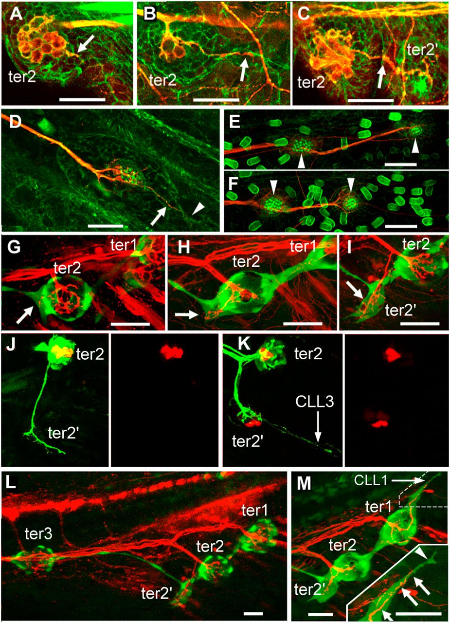
Fig. 3 Development of the terminal and caudal systems. (A–F) terminal and caudal budding in Thunnus simultaneously labeled for actin by phalloidin labeling and for acetylated tubulin by immunolabeling. (A and B) Neurite extension from ter2 (arrow) prefigures the formation of bud-neuromast ter22 in Thunnus. (C) ter2′ has formed and remains neurally connected to ter2 (arrow). (D) First neuromast of a caudal line with a neurite branch (arrow) extending into a budding extension (arrowhead). (E and F) First two neuromasts (arrowheads) of the two dorsalmost CLL lines of the same larva, showing that the caudal lines form by sequential budding. (G–M) Terminal and caudal budding in Danio. (G–I) Ventral migration of ter2 and extension of a neurite in the direction where ter2′ will form (arrows), as visualized in 12-dpf nbt-dsred; ET20 fish. (J and K) Formation of hair cell precursors (red) takes place only in the budding structure at 12–13 dpf, after the neurite (green) has sent a branch to presumptive tail line CLL3, as visualized in Hgn39d; atoh-tomato fish. (L) Overall pattern of ter neuromasts when bud-neuromast ter2′ appears, as visualized in nbt-dsred; ET20 fish. (M) Onset of the budding of tail line CLL1 from ter1. (Inset) Higher magnification of the dash-outlined area. A neurite (arrows) extends along the cell process (arrowhead) that emanates from ter1 to initiate CLL1. (Scale bars in A–M: 20 μm.)