
|
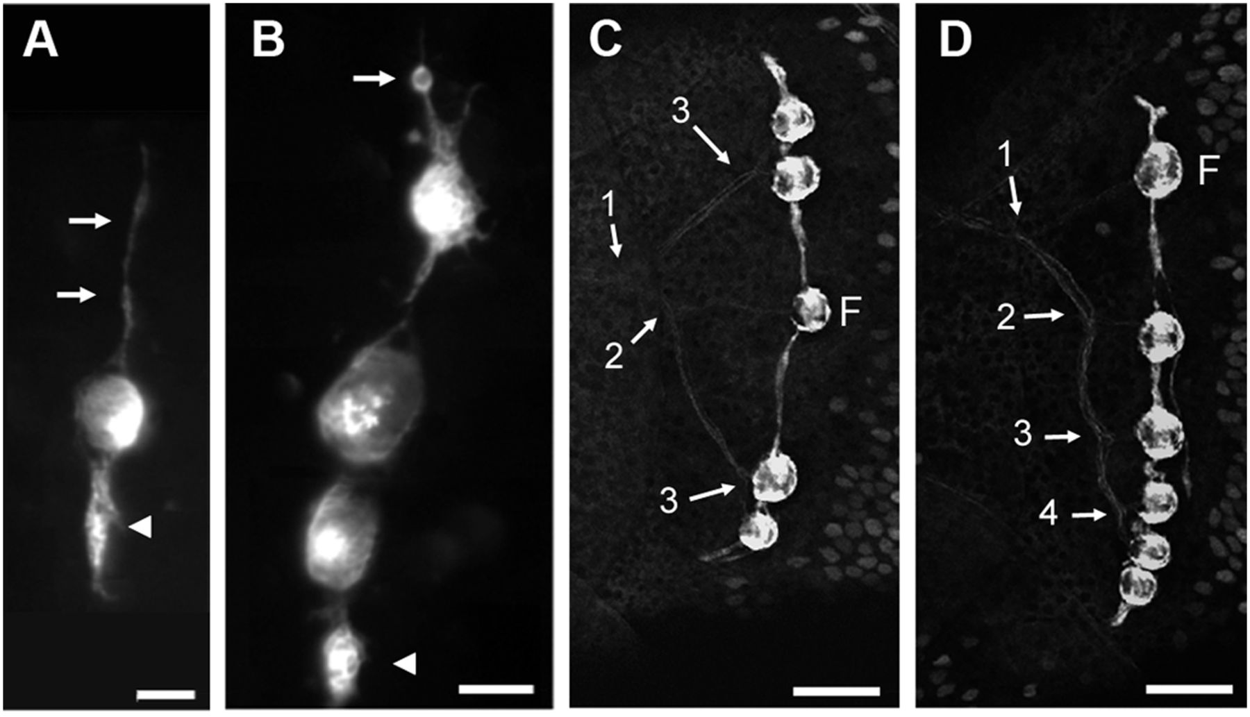
Fig. 1 Stitches in zebrafish. (A and B) Initiation of stitch formation in cxcr4.139:rfp fish. Long cell processes extend from the founder neuromast along the dorsoventral axis (A, arrows) and progressively swell up (arrowhead). Cell proliferation (B, arrow: cell rounding up before mitosis) eventually leads to the formation of a new, complete accessory neuromasts (B, arrowhead). (C and D) Innervation of stitches in Hgn39d; ET20 fish. The branching pattern reveals the history of stitch formation. Numbers refer to the successive branching/budding events. F: founder neuromast. In all figures, anterior is left and dorsal is up. (Scale bars: A and B—20 μm; C and D—50 μm.)