
|
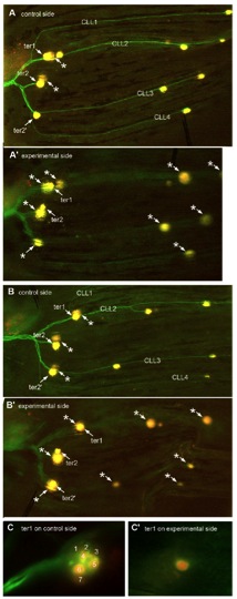
Fig. S4 Caudal fin lines do not form in the absence of innervation as visualized in Hgn39d fishes where the hair cells are labeled with DiAsp; asterisks mark out-of-focus neuromasts on the contralateral side. (A) ter2′ and all four caudal lateral lines (CLL) lines are present on the control side of 40-dpf fish, but not after complete ganglion ablation at 5 dpf on the experimental side (A′). (B) Even when ter2′ is present, due to ganglion ablation being completed only at 10 dpf, all CLL lines are still absent on the experimental side (B′). (C) A case of extreme reduction in the number of labeled hair cells in a denervated ter1 neuromast. At least seven hair cells were labeled on the control side (C), and only one was seen on the experimental side (C′). In A and B, ablation was performed on the left ganglion; both sides were photographed independently, and the figure showing the control side was subsequently inverted to facilitate comparison with the experimental side. A and B are assembled from consecutive frames of Z-stacks; C shows single frames.