
|
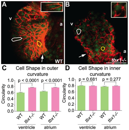
Fig. 2 Cell shape defects in tbx1-/- mutants.
(A, B) Confocal projections of hearts from Tg(cmlc2:EGFP) embryos at 48 hpf stained with Alcama antibody (red) to demarcate cell boundaries. Cells in the outer and inner chambers are outlined in white and yellow respectively. The insets in E and F show a magnified view of the outer curvature cell outlined in white. The plot in C shows that cells in the outer chamber of tbx1-/- mutants are rounder (circularity tending towards 1) as compared to WT. (D) Cell shape is unchanged in the inner chamber of tbx1-/- mutants. Each bar in plots C and D represents data collected from 70 cells in 7 different embryos. Arrows point to the AVC; v, ventricle; a, atrium. Scale bars: 25 μm.