
Fig. 1
- ID
- ZDB-IMAGE-130426-12
- Genes
- Publication
- Dooley et al., 2013 - On the embryonic origin of adult melanophores: the role of ErbB and Kit signalling in establishing melanophore stem cells in zebrafish
- All Figures
- Figures for Dooley et al., 2013
|
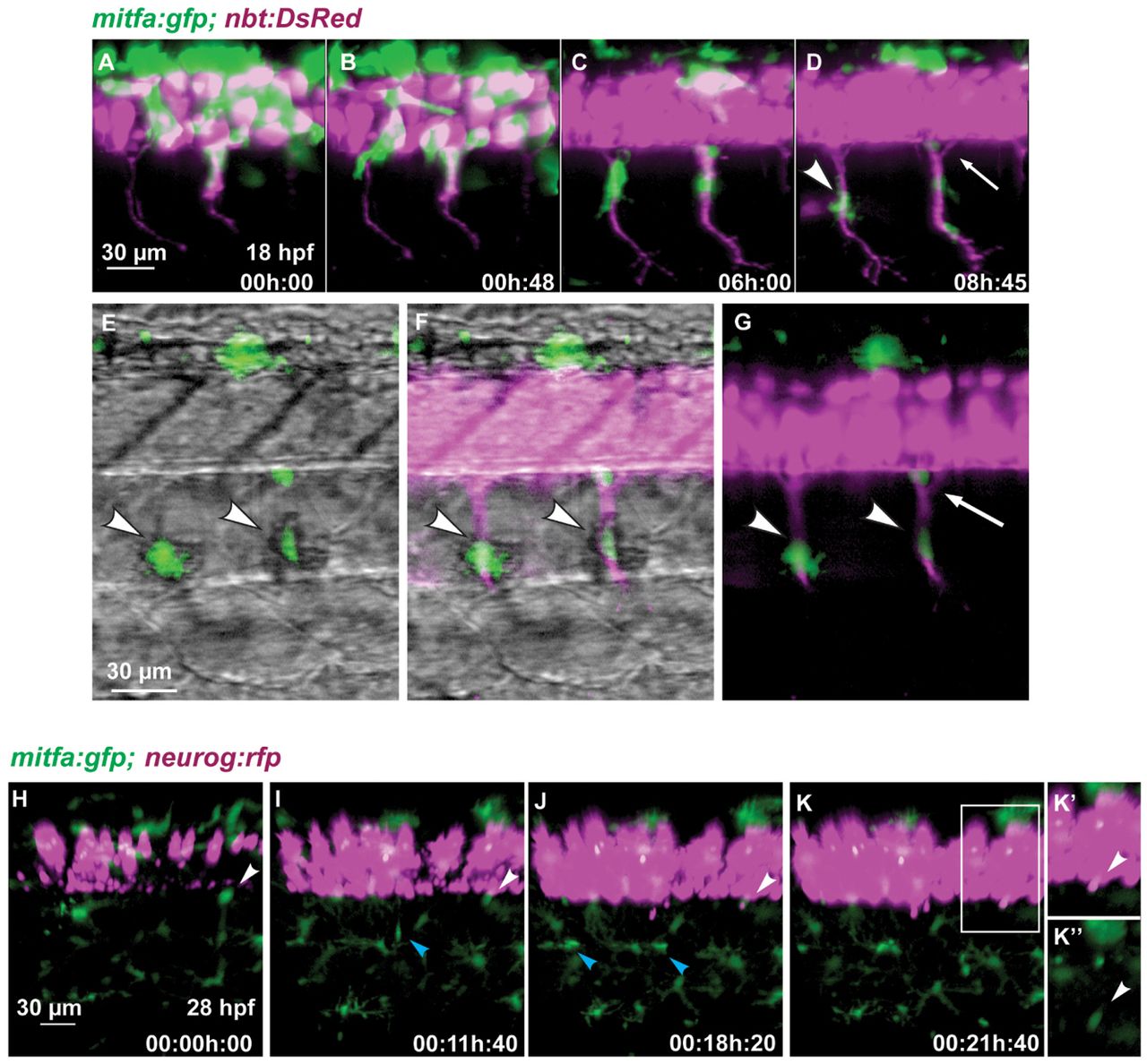
Fig. 1 Melanoblasts migrate along the motor neurons. (A-G) Time-lapse confocal imaging of a Tg(mitfa:gfp;nbt:DsRed) zebrafish embryo. Medially migrating GFP-positive cells (arrowhead in D) migrate along ventrally extending primary motor axons (DsRed) starting at 18 hpf. A portion of the area in D is shown in G, and in E,F with brightfield illumination. Arrowheads in E-G indicate melanoblasts melanising in situ. A GFP-positive cell remains at a ventral position of the neural tube (arrow in D,G). (H-K′′)Tg(mitfa:gfp;-8.4neurog1:nrfp) embryo imaged starting at 28 hpf over a period of 22 hours. A GFP-positive cell (white arrowheads) located close to the ventral base of the neural tube remains at this position over the next 21 hours when the expression of nRFP marks the appearance of the DRG in the same region. A medially positioned cell (blue arrowhead in I) migrates out to the horizontal myoseptum and divides to form melanophores of the lateral stripe (blue arrowheads in J). (K′) Enlargement of the boxed region in K. (K′′) Green channel only.