
|
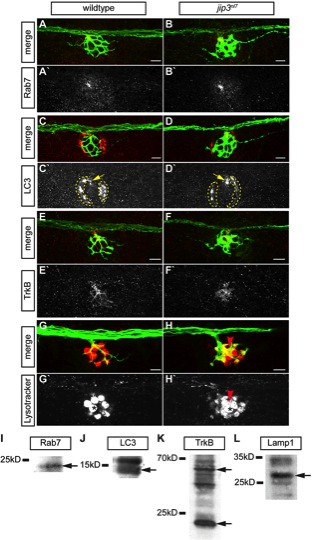
Fig. S3 Lysosomes, but not late endosomes, signaling endosomes or autophagosomes, accumulated in jip3nl7 axon terminals. (A–H) The density of late endosomes, autophagosomes, TrkB, and lysosomes were assayed in wildtype and jip3nl7 axon terminals by immunolabeling (A–F) and live imaging (G,H). (A,B) Rab7, a marker of late endosomes, was unchanged in jip3nl7 axon terminals. (C,D) LC3, a marker of autophagosomes, was unchanged in jip3nl7 axon terminals (yellow arrow). Note the high LC3 expression in NM support cells (dotted outline). (E,F) TrkB levels were slightly decreased in jip3nl7 axon terminals. (G,H) Lysotracker red staining at 5 dpf revealed elevated levels of lysosomes in jip3nl7 axon terminals (red arrowhead). Asterisk points out representative hair cells. This cell type has high Lysotracker labeling due to the large number of acidic vesicles. (I–L) Western blot analyses of 3 dpf embryo lysates demonstrate the specificity of the antibodies used. Arrows indicate bands corresponding to those that match the size of the predicted zebrafish protein orthologs.