
|
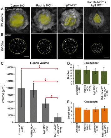
Fig. 5 Genetic interaction between Lgl2 and Rab11a regulates KV lumen formation uncoupled from ciliogenesis. (A,B) Images of GFP-labeled KV cells were used to determine KV lumen volumes (pseudocolored yellow) in live Tg(sox17:GFP) embryos (A) and acetylated tubulin antibodies were used to stain KV cilia (B) in MO control embryos and embryos injected with sub-optimal Rab11a MOlow, Lgl2 MOlow or co-injected with Rab11a MOlow + Lgl2 MOlow. The dashed circle outlines the approximate boundary of KV lumen. Scale bars: 20 μm. (C) Average volume of KV lumen at the 8-somite stage. (D,E) Analysis of KV cilia number (D) and length (E) showed that KV cilia were not significantly affected in Rab11a MOlow + Lgl2 MOlow embryos. Error bars indicate s.d. n, number of embryos analyzed. *P<0.05.