
|
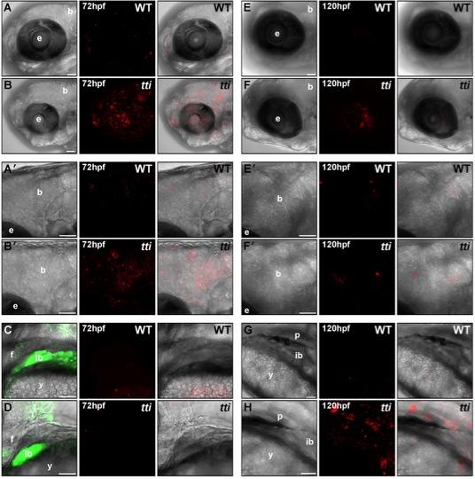
Fig. S4 LC3II-containing autophagosomes are found in multiple tissues in ttis450 larvae at 72 hpf and 120 hpf. (A–H) RNA encoding a mCherry-LC3 fusion protein was injected into the yolk of 1–4 cell zebrafish embryos derived from a pairwise mating of ttis450/+ heterozygotes (on the gutGFP background) and allowed to develop until the indicated time-point in the presence of chloroquine for the final 14 h. Maximum intensity projection images of a z series of confocal sections through WT [A, A′ (boxed area in A), C, E, E′ (boxed area in E) and G] and ttis450 larvae [B, B′ (boxed area in B), D, F, F′ (boxed area in F) and H] showing accumulated autophagosomes (red puncta) in the brain, eye and digestive organs (marked by GFP fluorescence in C, D) at 72 hpf (A–D) and 120 hpf (E–H). Scale bars = 50 μM. b, brain; e, eye; ib, intestinal bulb; f, fin; y, yolk; p, pancreas.