
|
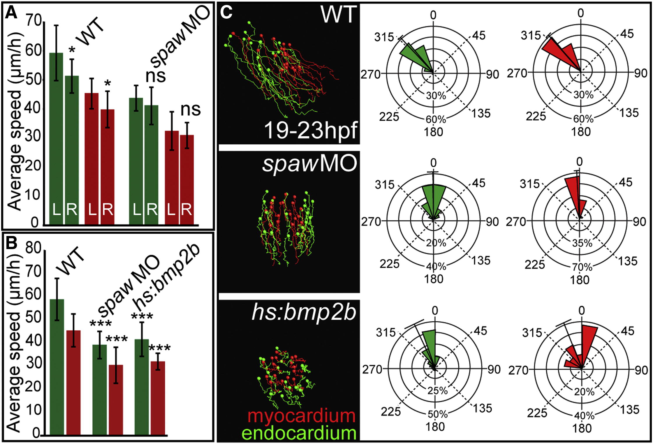
Fig. 3 Nodal and Bmp Signaling Affect Cardiac Progenitor Cell Motility and Laterality(A) Quantifications of cell motilities based on time-lapse analyses of endothelial and myocardial reporter lines between 19 and 23 hpf. In WT, endocardial (green) and myocardial (red) progenitor cell motility rates are significantly different between the left and right (mean with SD based on quantifications of at least n = 22 endocardial and n = 30 myocardial cells per genotype; p < 0.025), whereas no significant L/R differences are observed in spaw morphants.(B) Average speed measurements for spaw morphants or Bmp2b-overexpressing endocardial (green) and myocardial (red) progenitor cells reveal highly significant differences compared with WT (mean with SD based on quantifications of at least n = 16 endocardial cells and n = 16 myocardial cells per genotype; p < 0.0005).(C) Myocardial (red) and endocardial (green) progenitor cell tracks based on live imaging. Bearing angles summarize all endocardial and myocardial cell tracks. ns, not significant.See also Movies S3 and S4.
Reprinted from Developmental Cell, 24(6), Veerkamp, J., Rudolph, F., Cseresnyes, Z., Priller, F., Otten, C., Renz, M., Schaefer, L., and Abdelilah-Seyfried, S., Unilateral dampening of bmp activity by nodal generates cardiac left-right asymmetry, 660-667, Copyright (2013) with permission from Elsevier. Full text @ Dev. Cell