
|
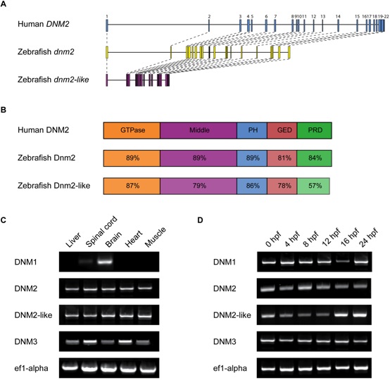
Fig. 2 Structure and expression of dnm2 and dnm2-like.(A) Molecular intron-exon organization of human DNM2, zebrafish dnm2 and zebrafish dnm2-like. (B) Protein structure of zebrafish Dnm2 and Dnm2-like compared to human DNM2. Percent identity between zebrafish and human protein domains was calculated using BLASTP. PH, pleckstrin homology domain; GED, GTPase effector domain; PRD, proline-rich domain. (C) RT-PCR was used to assay spatial expression levels of dnm2 and dnm2-like in tissues isolated from adult zebrafish. Primers for ef1α were used as an internal control. (D) RT-PCR was used to assay temporal expression levels of dnm2 and dnm2-like between 0 hpf and 24 hpf. All classical dynamins appear to be deposited as maternal mRNAs and expressed throughout early development.