
|
Fig. 6
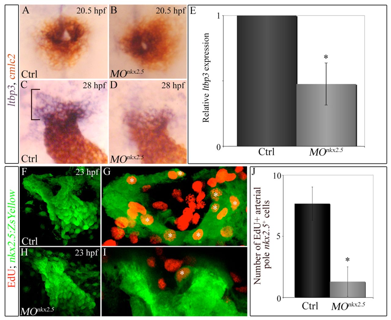
Knocking down nkx2.5 compromises arterial-pole ltbp3 expression and SHF proliferation. (A-D) Double in situ hybridization of cmlc2 and ltbp3 transcripts in control (A,C) and morphant (B,D) embryos at 20.5 (23-somite stage, cardiac cone stage; n=20, control; n=32, morphant) and 28 (linear heart tube stage; n=20, control; n=30/48 morphants) hpf. Bracket highlights non-myocardial ltbp3+ SHF progenitors. (E) Graph showing the average relative abundance of ltbp3 transcripts in morphant compared with control embryos at 28 hpf determined by quantitative PCR. Error bar represents one s.d., *P<0.05. (F-J) nkx2.5 morphants exhibit reductions in proliferating arterial-pole nkx2.5+ SHF cells. At 23 hpf, control and morphant Tg(nkx2.5:ZsYellow) embryos were double immunostained for incorporated EdU (red) and ZsYellow protein (green). Confocal z-stack images of heart tube regions in control (F,G) and morphant (H,I) embryos captured through green and red filters. (F,H) Flattened z-stack images (green channel only) of representative control (F) and morphant (H) heart tubes. (G,I) Arterial poles of heart tubes shown in F and H. Three contiguous confocal slices (red and green channels merged) from F and H were flattened. White asterisks mark double-positive cells (EdU+ red nuclei surrounded by ZsYellow+ green cellular fluorescence). (H) Average numbers of double-positive cells in control and morphant heart tubes. n=6 embryos per experimental group. Error bars represent one s.d., *P<0.0001. hpf, hours post-fertilization; Ctrl, control; MO, morpholino.