
Image
|
Figure Caption
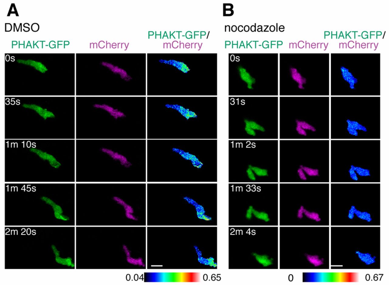
Fig. 4 Microtubule disassembly inhibits PI(3)K activation at the leading edge. (A) Time-lapse ratiometric imaging (PHAKT-GFP/mCherry) of PI(3,4,5)P3–PI(3,4)P2 dynamics during neutrophil random motility in the head. (B) Microtubule depolymerization inhibits localization of PI(3,4,5)P3–PI(3,4)P2 at the leading edge. Scale bars: 10 μm.
Acknowledgments
This image is the copyrighted work of the attributed author or publisher, and
ZFIN has permission only to display this image to its users.
Additional permissions should be obtained from the applicable author or publisher of the image.
Full text @ J. Cell Sci.