
|
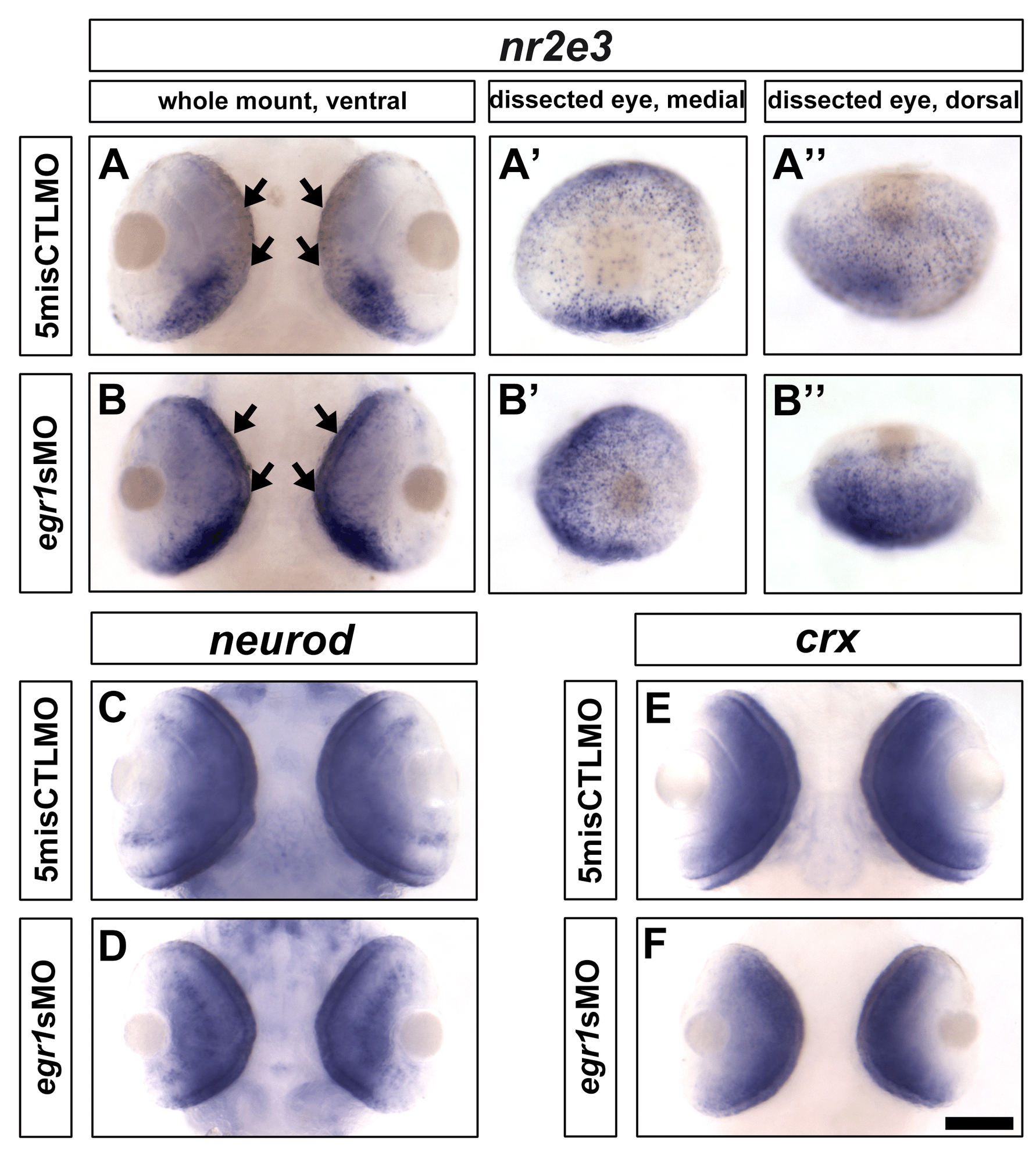
Fig. S3
In situ hybridization of nr2e3, neurod and crx at 72 hpf. (A & B) The staining of nr2e3 in the Egr1-morphant retinas was higher from the ventral (B) and dorsal (B′′) views compared with the controls (A & A′′). From the medial view, the PRs that were stained as individual dots were widely distributed in the Egr1-morphant retinas (B′), while they were relatively sparse in the control retinas, especially in the central region (A′). For neurod and crx, their expression patterns and levels were comparable between the control (C & E) and Egr1-morphant (E & F) retinas. Thus, these observations suggest that egr1 negatively regulates nr2e3 but not neurod and crx at 72 hpf. Nonetheless, since PRs ultimately differentiated relatively normally in the Egr1 morphants at 120 hpf (Figure 7), the results are more consistent with the possibility that the development of PRs was delayed in the morphants. Scale bar = 100 µm.