
|
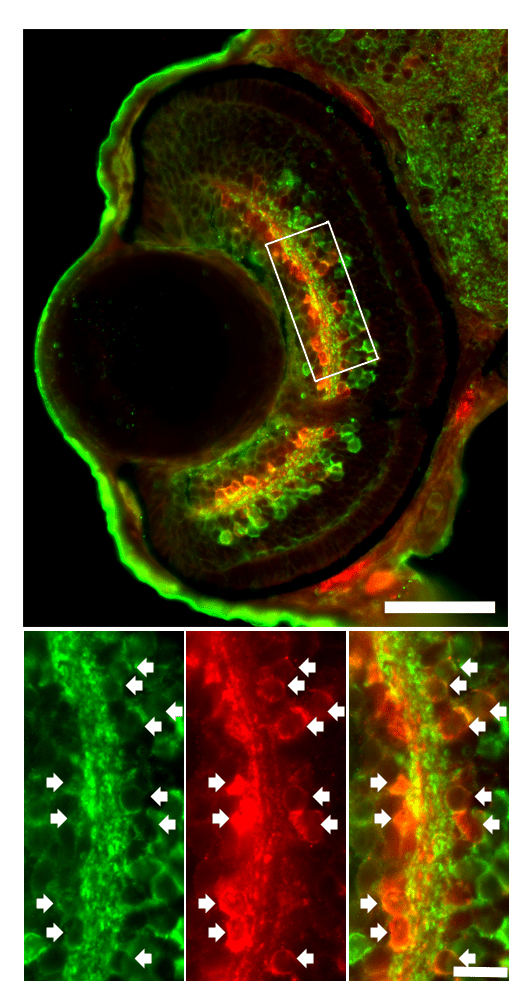
Fig. S1 Amacrine cells immunolabeled by Parv and GABA markers. (Top) An overlay image of GABA+ (green) and Parv+ (red) cells in a normal WT retina at 72 hpf. (Bottom) A magnified view of the white box at the top. From left to right: GABA, Parv and the overlay image. Many of the Parv+ AC cell bodies were also GABA+ (white arrows), suggesting they might be a subset of GABAergic ACs. Note that there were overlapping and non-overlapping GABA+ and Parv+ regions in the IPL, suggesting that these ACs projected to different sub-laminae in the IPL. Scale bar = 50 µm for the top image and 10 µm for the bottom images.