
|
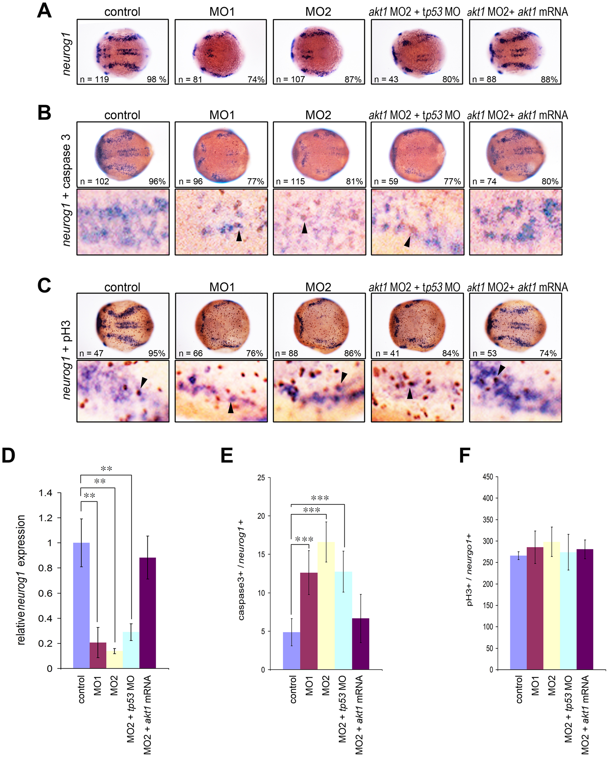
Fig. 3 Decreased neuronal precursors in Akt1 knockdown embryos during early neurogenesis.
(A) In situ hybridization showing the dramatic downregulation of neurogenin1 in akt1 morpholino-injected embryos. This effect was rescued by co-injection with akt1 mRNA but not a tp53 morpholino, which was confirmed by qPCR analysis (D). (B) Apoptotic cells were detected using caspase-3 antibody (brown) and double-labeled with neurogenin1 (purple), which showed an increase in apoptotic cells in akt1 morphants. The double-labeled cells are indicated with arrows and quantified in E. (C) Proliferating neurons were double-labeled with phosphohistone H3 antibody and neurogenin1 riboprobes (arrows), which showed no significant difference between akt1 morphants and controls. This was confirmed by quantification (F). In A–C the lower panels are enlargements of the upper panels. **, p<0.01; ***, p<0.001.