
|
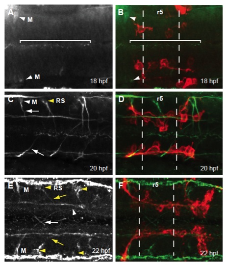
Fig. S1 Early FBMN migration into r5 and r6 precedes both Mauthner and reticulospinal neuron axon projection into the hindbrain. Maximum projection dorsal views of Tg(zCREST:membRFP) embryos immunostained with an acetylated tubulin antibody (A,C,E) and merged images (green; B,D,F). (A,B) At 18 hpf, Mauthner (M) neuron cell bodies are present in r4 (white arrowhead) but their axons are not yet present. Cilia in the lumen of the hindbrain (bracket) as well as stereocilia in the otic vesicle (not shown) are present at this stage, verifying antibody staining efficacy. (C,D) At 20 hpf, axons of the Mauthner neurons have begun to project towards the hindbrain midline (white arrow). Additionally, reticulospinal (RS) neuron cell bodies are present (yellow arrowhead) and have begun to project their axons toward the MLF. (E,F) At 22 hpf the axons of the Mauthner neurons have crossed at the midline in r5 (white arrow) and will soon contribute to the MLF axon tract (white arrowhead). RS neurons (yellow arrowhead) have sent out projections that have contributed to the MLF (yellow arrow).