
|
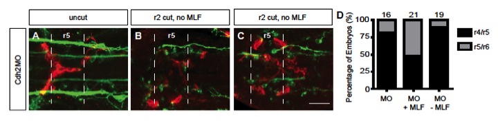
Fig. S4 Loss of both Cdh2 and MLF axons in the hindbrain blocks FBMN migration. (A-C) Maximum projection dorsal views of FBMNs (red) and the MLF (green) in 24 hpf Cdh2-depleted Tg(zCREST1:membRFP) zebrafish embryos stained with zn-12 antibody. In all treatments, FBMNs coalesce in the midline; however, FBMNs never migrate along either the LLF or projections of the reticulospinal neurons. Broken lines highlight r5 boundaries. (A) Uncut control Cdh2-depleted embryos at 24 hpf show typical Cdh2-depletion phenotype of FBMN migration into r5 and coalescence of neurons in the midline. In a smaller proportion of Cdh2-depleted embryos, FBMNs still coalesce at the midline but are able to migrate into r6. (B) In embryos lacking both Cdh2 and the MLF, the majority of embryos show FBMNs that migrate only into r5. (C) In a smaller number of embryos lacking both Cdh2 and the MLF, FBMNs migrate into r6. (D) Percentage of embryos affected by Cdh2 depletion and severing manipulations. Cdh2 depletion alone (MO) results in 82% of embryos exhibiting stalled FBMN migration in r5 and 18% with FBMNs migrating into r6. In Cdh2-depleted embryos in which the embryos were severed incompletely so the MLF is still present (MO +MLF), 48% of embryos show stalled migration in r5, whereas 52% of embryos show FBMN migration reaching r6. In embryos depleted of both Cdh2 and the MLF (MO –MLF), 89% result in FBMNs stalling in r5 and 11% result in FBMNs reaching r6. The number of embryos scored is indicated. Scale bar: 20 μm.