
|
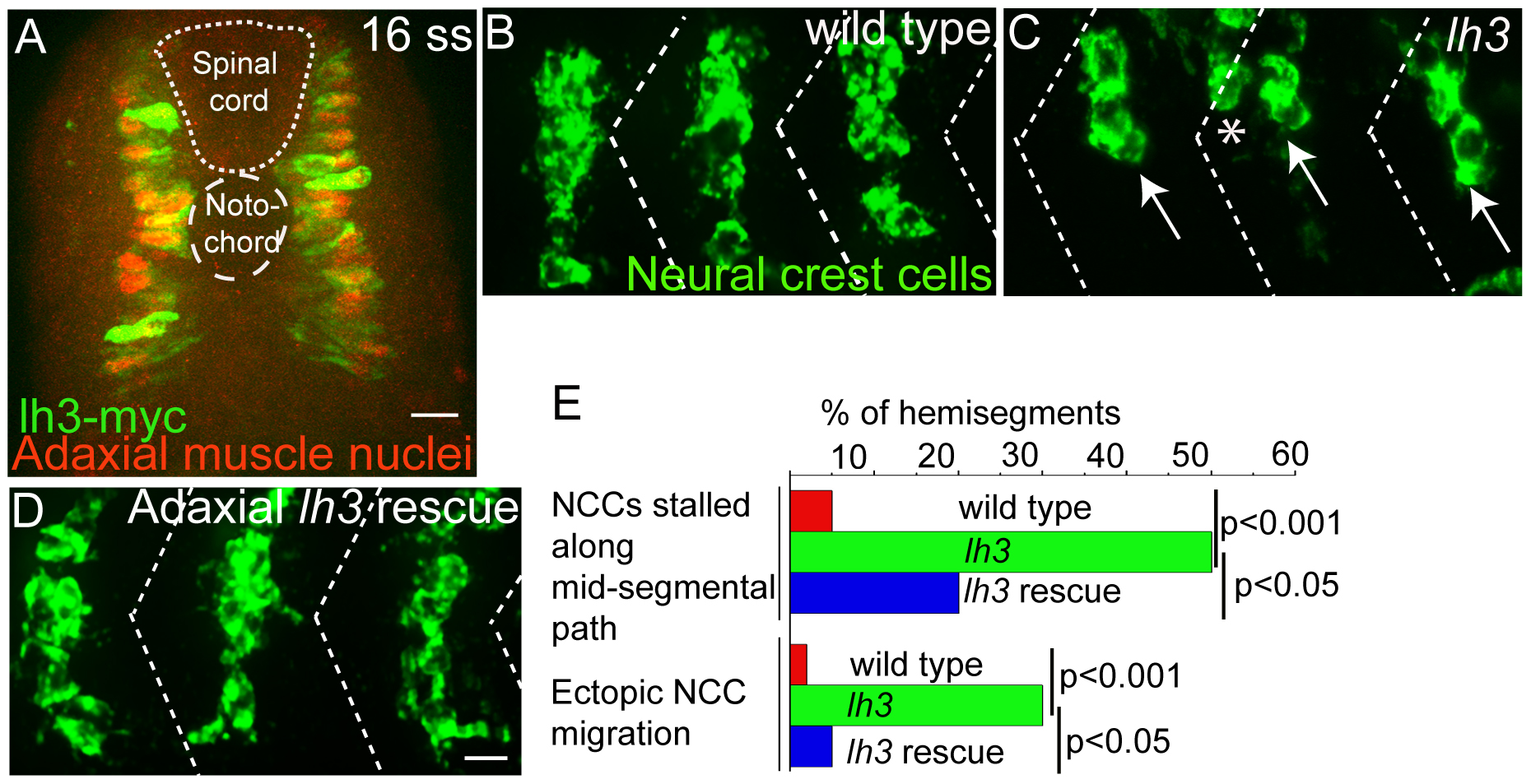
Fig. 3 Lh3 activity in adaxial cells can rescue neural crest cell migration defects in lh3 mutants
. Cross sectional view of 16 somite stage [Tg(smhc1:lh3-myc)] embryos showing expression of lh3-myc transgene (green) in prox-1 (red) positive adaxial cells. Lateral view of an 28 hour old lh3 sibling embryo (B), lh3 mutant (C) and an lh3 mutant embryo expressing lh3-myc in adaxial muscle cells (D), all stained with crestin (green). Note the stalled neural crest cells in the mid-segmental and neural crest cells in the hemisegment region in lh3 mutants (arrows and asterisks in C). These defects were rescued in by expressing lh3-myc in adaxial cells of lh3 mutants (C). (E) Quantification of neural crest cell migration defects. P values (** p<0.001; * p<0.05) were obtained using Chi- Square test. Scale bar-10 micron.