
|
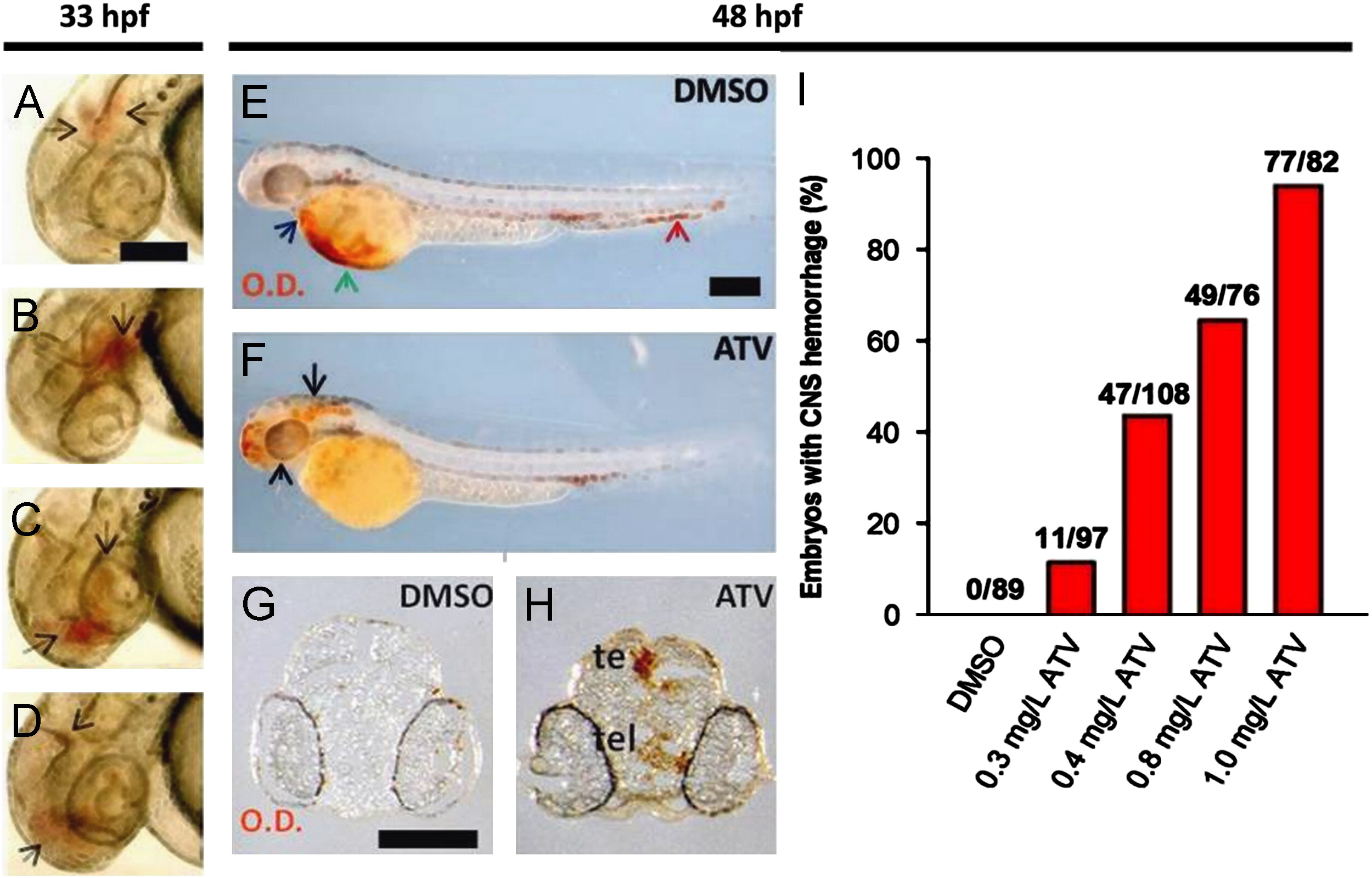
Fig. 2 Pharmacological inhibition of the HMGCR pathway induces cerebral hemorrhage in developing zebrafish. ((A)–(D)) Photomicrographs of embryos soaked in 0.5 mg/L ATV and imaged at 33 hpf. Black arrows denote areas of hemorrhage. Scale bar=200 μm. ((E) and (F)) Bright-field images of zebrafish embryos soaked in DMSO or 0.5 mg/L ATV, stained with o-Dianisidine (OD) and imaged at 48 hpf. Black arrows indicate sites where abnormal accumulation of hemoglobinized blood was detected in the brain. The blue arrow denotes OD-positive cells in the pericardium, the green arrow shows the sinus venosus and the red arrow designates the tail region. Scale bar=200 μm. ((G) and (H)) Transverse sections through embryo heads at 48 hpf stained with OD. Abbreviations: te, tectum; tel, telencephalon. Scale bar=200 μm. (I) Percentage of embryos treated with various doses of ATV and scored for the presence of cerebral hemorrhage at 48 hpf. Numbers above the bars represent the ratios used to calculate the percentages.
Reprinted from Developmental Biology, 373(2), Eisa-Beygi, S., Hatch, G., Noble, S., Ekker, M., and Moon, T.W., The 3-hydroxy-3-methylglutaryl-CoA reductase (HMGCR) pathway regulates developmental cerebral-vascular stability via prenylation-dependent signalling pathway, 258-266, Copyright (2013) with permission from Elsevier. Full text @ Dev. Biol.