
|
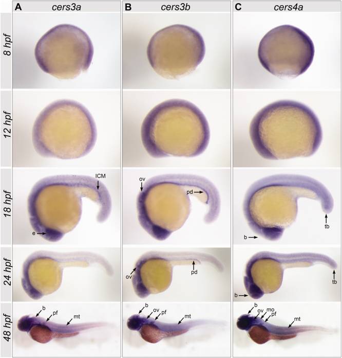
Fig. 3 Spatial expression patterns of zebrafish cers3a, cers3b, and cers4a in gastrula (8 hours postfertilization [hpf]), 6-somite stage (12 hpf), 18-somite stage (18 hpf), 24 hpf, and 48 hpf embryos. A: The cers3a pattern at 18 hpf includes the eye (e) and intermediate cell mass (ICM) and a subset of putatively neuronal cells. At 48 hpf, an expression is found in the brain (b), pectoral fin (pf), and myotomes (mt). B: cers3b expression is found in the otic vesicle (ov) and pronephric duct (pd) at 18 hpf and 24hpf. At 48 hpf, cers3b is detected in the brain (b), otic vesicle (ov), pectoral fin (pf), and myotome (mt). C: cers4a shows an ubiquitous pattern in all stages analyzed, most pronounced in the brain (b) and tail bud (tb). At 48 hpf, the expression is particularly localized in brain (b), otic vesicle (ov), medulla oblongata (mo), pectoral fin (pf), and myotomes (mt).