
Image
|
Figure Caption
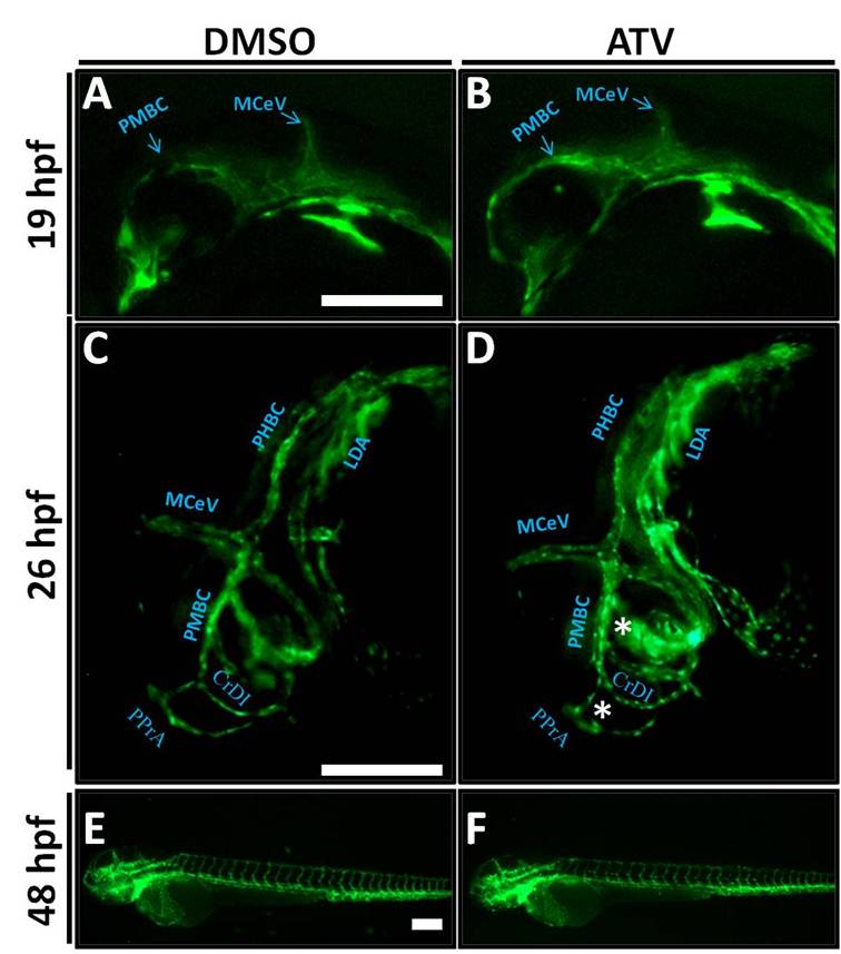
Fig. S2 Embryos with cerebral hemorrhage have normal vascular patterning and no regression. (A-F) Representative photomicrographs of Tg(fli1:EGFP) embryos exposed to DMSO or 0.5 mg/L ATV and imaged at 14 hpf (A, B), 26 hpf (C, D) and 48 hpf (E, F). White asterisks indicate abnormally dilated PPrA and PMBC. Anterior is to the left and dorsal to the top. Scale bars = 200 μm.
Figure Data
Acknowledgments
This image is the copyrighted work of the attributed author or publisher, and
ZFIN has permission only to display this image to its users.
Additional permissions should be obtained from the applicable author or publisher of the image.
Reprinted from Developmental Biology, 373(2), Eisa-Beygi, S., Hatch, G., Noble, S., Ekker, M., and Moon, T.W., The 3-hydroxy-3-methylglutaryl-CoA reductase (HMGCR) pathway regulates developmental cerebral-vascular stability via prenylation-dependent signalling pathway, 258-266, Copyright (2013) with permission from Elsevier. Full text @ Dev. Biol.