
|
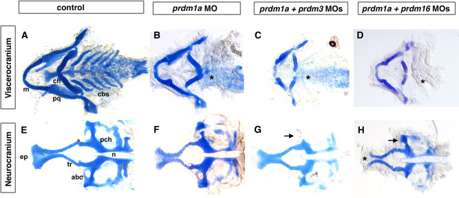
Fig. 4 Combination of sub-optimal doses of prdm1a with prdm3 or prdm16 Morpholino results in severe craniofacial defects. Alcian blue staining on 5-dpf larvae to detect cartilage formation. Alcian blue–stained uninjected (A, E), prdm1a (B, F), prdm3, prdm1a-prdm3 double (C, G), and prdm1a-prdm16 double (D, H) morphant larvae. As compared to control (A), prdm1a knockdown alone has an inverted ceratohyal (ch) and missing ceratobranchial 2-5 (*) in the viscerocranium (B). In addition, prdm1a morphants have a smaller and narrower neurocranium and slightly shortened trabeculae (F). The combination of sub-optimal doses of prdm1a with prdm3 e3i3 Morpholino resulted in a more severe phenotype in viscerocranium (C, shortening of the Meckel′s cartilage and loss of ceratobranchial 2–5). In the neurocranium, the double morphants have an increasingly smaller and thinner neurocranium, a smaller ethmoid plate, and shorter trabeculae. The combination of suboptimal doses of prdm1a and prdm16 ATG Morpholino led to a more severe phenotype in the neurocranium (H, much smaller and thinner neurocranium with clefting observed in 20-30% of embryos, smaller ethmoid plate, shorter trabeculae) and viscerocranium (D, shortening of the Meckel′s cartilage and absence of ceratobranchial 2–5). Anterior is to the left. abc, anterior basicapsular commissure; cbs, ceratobranchials; ch, ceratohyal; ep, ethmoid plate; m, Meckel′s cartilage; n, notochord; pch, parachordal; tr, trabeculae; * in B–D illustrates missing cbs, in H indicates area of clefting.