
|
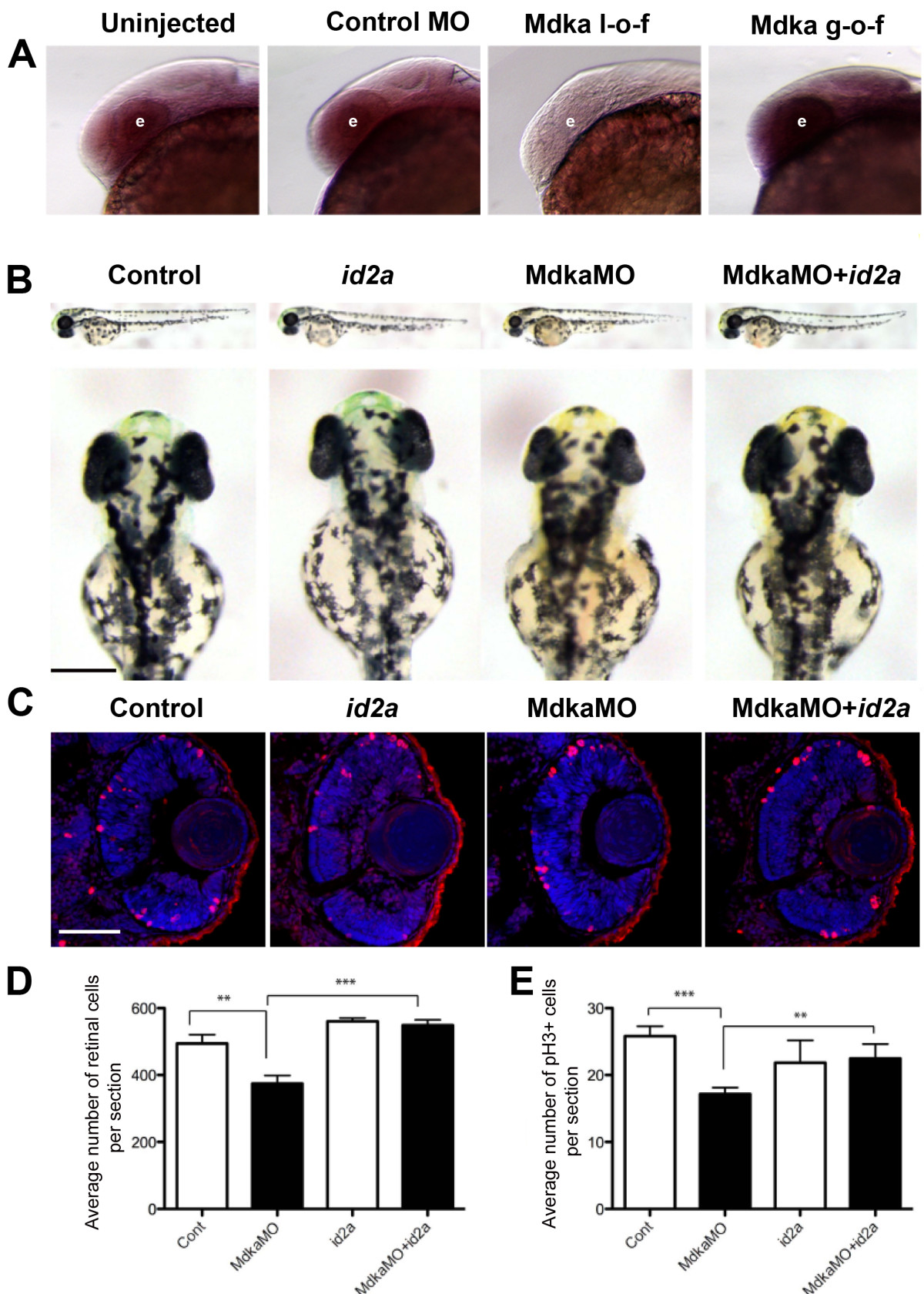
Fig. 6 Mdka functions upstream of Id2a. (A) Wholemount in situ hybridizations showing id2a expression at 28 hpf. Embryos were uninjected (Uninjected), injected with mismatch Mdka morpholinos (Control MO), Mdka translation-blocking morpholinos (Mdka l-o-f), or transgenic embryos treated with heat shock at 24 hpf (Mdka g-o-f). The letter ‘e’ identifies the eye. Note that id2a expression is modulated by Mdka levels. (B) Representative control and experimental embryos at 48 hpf. Embryos were uninjected (Control), injected with id2a mRNA (id2a), Mdka translation-blocking morpholinos (MdkaMO), or co-injected with Mdka translation-blocking morpholinos and id2a mRNA. (C) Representative retinal sections from embryos at 48 hpf. The conventions are as above. Sections are stained with DAPI to label nuclei and antibodies against phosphohistone H3. (D) Graph showing the number of retinal cells in each group. (E) Graph showing the number of phosphohistone H3-positive cells in each group. Note that the number of DAPI and phosphohistone H3-positive cells are reduced following Mdka l-o-f and cell numbers are restored following co-injections of Mdka translation-blocking morpholinos and id2a mRNA. **P <0.05. Scale bars in panel B and C are 200 μm and 50 μm, respectively.