
|
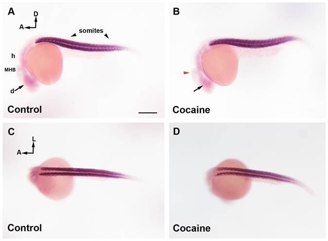
Fig. 5 miR-133b distribution in zebrafish embryos at 24 hpf by whole-mount ISH.
Control Group (A and C) and cocaine group (B and D). In a lateral view of miR-133b expression (A), the miRNA is mainly localized in somites and weakly in the brain (diencephalon, midbrain, MHB and hindbrain). Embryos exposed to cocaine (B) show a decrease in diencephalic expression (black arrow), and MHB (red arrowhead). A dorsal view of miR-133b (C) shows that this miRNA is mainly present in somites, although it is difficult to determine whether cocaine affects the expression of miR-133b in this area (D). Scale bars = 300 μm. A, B; C; D = 6X. A: anterior; D: dorsal; L: lateral; d: diencephalon; m: midbrain, h: hindbrain; MHB: Midbrain Hindbrain Boundary, sm: skeletal muscle.