
|
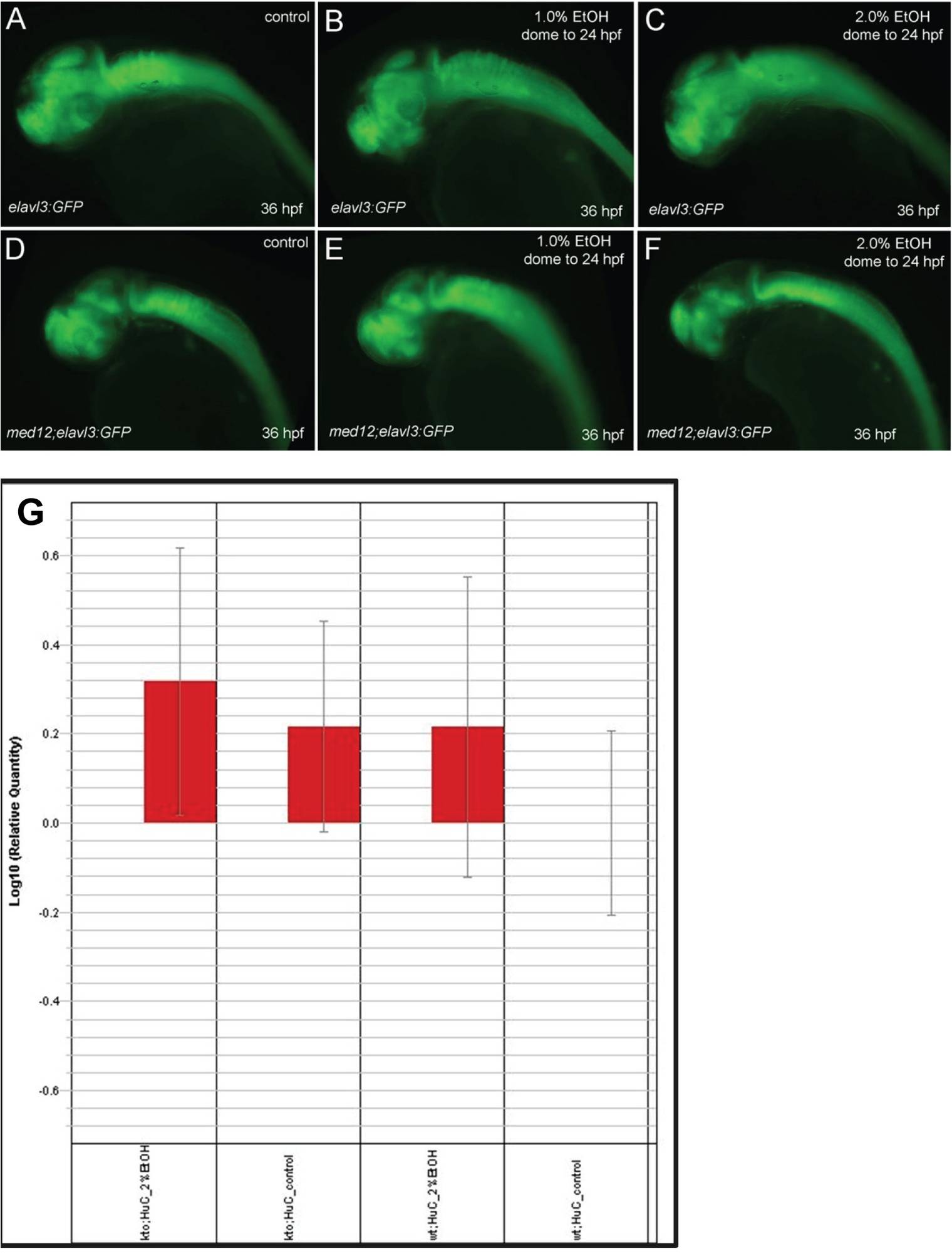
Fig. S5 Ethanol exposure had no effect on the overall numbers of post-mitotic neurons in wild-type Tg(elavl3:GFP) embryos, while med12y82 mutant embryos had fewer post-mitotic neurons that became further reduced after ethanol exposure. A-F, lateral view of 36 hpf embryos. A-C, wild-type Tg(elavl3:GFP) embryos. D-F, med12y82;Tg(elavl3:GFP) embryos. A, D, controls. B, E, treated with 1% ethanol from dome stage to 24 hpf. C, F, treated with 2% ethanol for 24 hrs at 3 dpf. G, quantitation of GFP expression by real-time quantitative RT-PCR. GFP expression was normalized to β-actin as an internal control. Relative quantity was compared to control wild-type Tg(elavl3:GFP) embryos (HuC). The relative quantity of GFP in control wild-type Tg(elavl3:GFP) embryos was set to one, which is zero on the log scale. The relative expression of GFP trends higher in med12y82 mutant (kto) embryos and in ethanol-treated embryos as shown by the red bars. Although the overall amount of GFP expression in med12y82 mutant and ethanol-treated embryos appears to be somewhat reduced in panels A-F, when normalized to β-actin GFP expression is essentially equal or even increased relative to control wild-type embryos. This suggests that neither ethanol exposure nor med12 mutation results in a relative reduction in post-mitotic neurons. Error bars represent 95% confidence levels.