
|
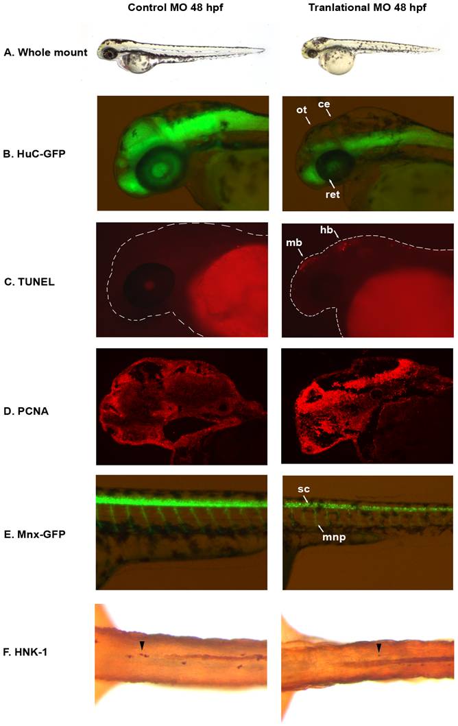
Fig. 2 auts2 48 hpf morphant phenotype.
(A) Fish injected with the 5 base-pair translational MO mismatch control have similar morphology as wild type fish. Injection of the auts2 translational MO results in fish with a stunted development phenotype that includes a smaller head, eyes, body and fins. (B) HuC-GFP fish injected with the 5 bp control MO display normal levels of developing neurons in the brain. HuC-GFP translational MO injected fish display considerably less developing neurons in the optic tectum (ot), retina (ret), and cerebellum (ce). (C) 5 bp mismatch control injected fish have little to non-observable apoptosis in the brain as observed by TUNEL staining, while translational MO injected fish display high levels of apoptosis, primarily in the midbrain (mb) and hindbrain (hb). (D) PCNA cell proliferation assay in the 5 bp MO control injected fish shows lower levels of cell proliferation in the brain compared to the translational MO injected fish. (E) Tg(mnx1:GFP) fish injected with the 5 bp MO control display normal levels of motor neurons versus the auts2 translational MO injected fish which have fewer motor neurons in the spinal cord (sc). In addition, motor neuron projections (mnp) are weaker and more perpendicular to the spinal cord. (F) Translational MO injected fish display fewer Rohon-Beard cells (arrowheads) in the spinal cord than morphants. All morphant fish are scaled to their 5 bp control counterparts.