
|
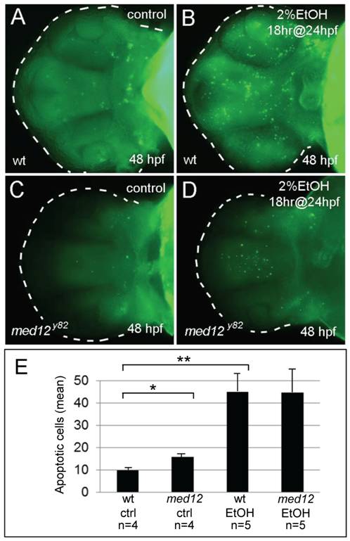
Fig. 5 Ethanol exposure causes similar levels of neuronal cell death in wild-type and med12 mutant embryos.
A-D, acridine orange stained embryos at 48 hpf, ventral views. A, untreated wild-type (wt) embryo. B, wt embryo treated with 2% ethanol for 18 hrs at 24 hpf. C, untreated med12y82 embryo. Med12y82 and wt embryos showed similar low levels of apoptosis. D, med12y82 embryo treated with 2% ethanol for 18 hrs at 24 hpf. Med12y82 and wt embryos treated with 2% ethanol showed similar increases in acridine orange staining. E, the average numbers of apoptotic cells in the brains of med12y82 and control embryos treated with ethanol as in panels AD. There were significantly more apoptotic cells in the brains of med12y82 embryos compared to wild-type embryos (p<0.05), however, following EtOH exposure, the numbers of apoptotic cells in the brains of both med12y82 and wild-type embryos greatly increased. The number of apoptotic cells in the brains of med12 mutant embryos increased 2.8-fold compared to their untreated med12 mutant siblings (p = 0.073), while apoptotic cells increased over 5-fold in ethanol-treated wild-type embryos compared to their untreated siblings (p<0.005). Error bars represent SEM. **p<0.01, *p<0.05.