
|
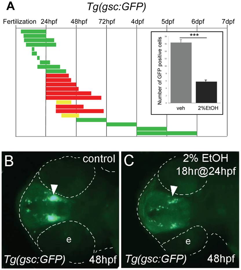
Fig. 1 Ethanol exposure reduces the number of gsc:GFP expressing neurons.
A, the duration and embryonic stage of 2% ethanol treatments in Tg(gsc:GFP) embryos are illustrated with solid bars. Colors indicate the treatment had no effect (green), slightly reduced (yellow), or reduced (red) GFP expression. Inset, the average numbers of GFP positive cells in the preoptic area (POA) of 72 hpf embryos treated with 2% ethanol for 24 hrs at 24 hpf (n = 12) and vehicle control (n = 10). Ethanol treatment resulted in a highly significant reduction of GFP-expressing cells in the POA, ***p<0.001. Error bars represent SEM. B, C, ventral views of Tg(gsc:GFP) embryos at 48 hpf. B, is the control. C, treated with 2% ethanol for 18 hrs at 24 hpf. GFP expression in the neuroendocrine preoptic area (arrowheads) was dramatically reduced in the embryos exposed to ethanol. The embryo and eyes (e) are outlined (dashed white line).