
|
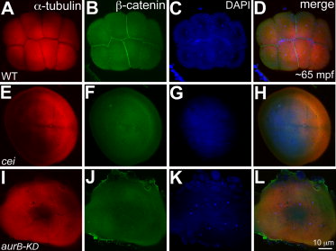
Fig. 3 Stage IV oocyte injection of aurB-KD mRNA phenocopies the cei/aurB maternal-effect phenotype. A–L: Animal views of 65 mpf blastodiscs immunolabeled to detect α-tubulin (A,E,I) and β-catenin (B,F,J), with DAPI (4′,6-diamidine-2-phenylidole-dihydrochloride) stainings (C,G,K) and panel merges (D,H,L). A,B: Robust furrows in wild-type embryos labeled with α-tubulin (A), which accumulate β-catenin (B). E,F: &alphja;-Tubulin labeling of control cei/aurB embryos from mutant stage IV oocytes show partial rudimentary furrows (E) with no β-catenin labeling (F). I,J: α-tubulin labeled partial, immature furrows in embryos from wild-type stage IV oocytes injected with aurB-KD mRNA (I), which also fail to accumulate β-catenin (J), recapitulating cei/aurB maternal effect phenotype. C,G,K: As in Figure 2, DAPI images show that nuclear division proceeds normally in all cases.