
|
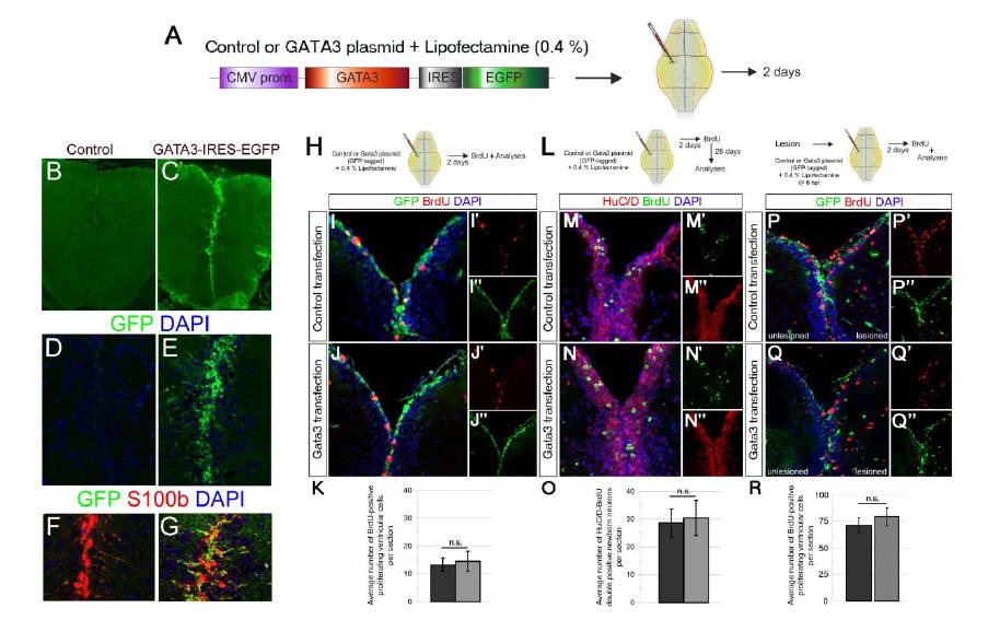
Fig. S3 Gata3 is not sufficient to induce regenerative neurogenesis without the lesion. (A) Schematics for lipofectamine-mediated plasmid transfection to ventricular cells of the adult zebrafish telencephalon. CMV promoter drives the expression of Gata3 open reading frame and GFP. Brains were analyzed at 2 days after cerebroventricular microinjection of the plasmids. (B) GFP immunohistochemical (IHC) staining on control plasmid (backbone vector)-transfected brains. (C) GFP IHC on Gata3-GFP plasmid-transfected brains. (D) Close-up to dorsomedial region of B. Counterstained with DAPI. (E) Close-up to dorsomedial region of C. Counterstained with DAPI. (F) GFP, S100b and DAPI staining on control-transfected brains. (G) GFP, S100b and DAPI staining on Gata3-transfected brains. Radial glial cells are effectively transfected. (H) After plasmid transfection, proliferating cells were detected with brief BrdU pulse for two hours before sacrificing the animals. (I) GFP, BrdU and DAPI staining on control-transfected brains. (I′) Individual channel for BrdU. (I′ ′) Individual channel for GFP. (J) GFP, BrdU and DAPI staining on Gata3-transfected brains. (J′) Individual channel for BrdU. (J′ ′) Individual channel for GFP. (K) Quantification graph for average number of BrdU-positive ventricular cells per section. Gata3 transfection does not induce cell proliferation. Bars represent ± s.e.m. (L) After plasmid transfection, a brief BrdU pulse for two hours was given and fish were sacrificed 28 days after BrdU treatment. (M) HuC/D, BrdU and DAPI staining on control-transfected brains. (M′) Individual channel for BrdU. (M′ ′) Individual channel for HuC/D. (N) HuC/D, BrdU and DAPI staining on Gata3-transfected brains. (N′) Individual channel for BrdU. (N′ ′) Individual channel for HuC/D. (O) Quantification graph for average number of newborn neurons per section. Gata3 transfection does not induce neurogenesis. Bars represent ± s.e.m. (P) GFP and BrdU immunostaining coupled to DAPI staining on 2 days post lesion (dpl) brains transfected with control plasmid at 6 hours post lesion (hpl). (P′) Individual channel for BrdU. (P′ ′) Individual channel for GFP. (Q) GFP and BrdU immunostaining coupled to DAPI staining on 2 days post lesion (dpl) brains transfected with Gata3 expression plasmid at 6 hours post lesion (hpl). (Q′) Individual channel for BrdU. (Q′′ ′) Individual channel for GFP. (R) Quantification graph for average number of BrdU-positive ventricular cells per section. Bars represent ± s.e.m. n = 6 fish for every experiment.
Reprinted from Developmental Cell, 23(6), Kizil, C., Kyritsis, N., Dudczig, S., Kroehne, V., Freudenreich, D., Kaslin, J., and Brand, M., Regenerative Neurogenesis from Neural Progenitor Cells Requires Injury-Induced Expression of Gata3, 1230-1237, Copyright (2012) with permission from Elsevier. Full text @ Dev. Cell