
|
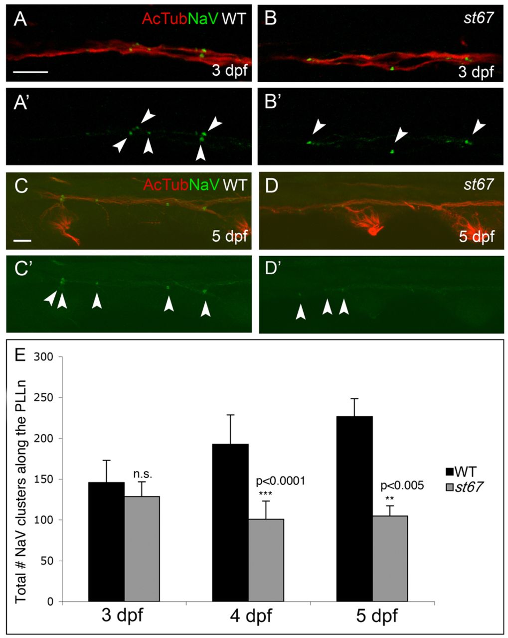
Fig. 1 Nodes of Ranvier are abnormal in st67 mutant zebrafish at 5 dpf. (A–D) Images of axons from the posterior lateral line nerve (PLLn) in larvae of the indicated genotypes at 3 (A,B) and 5 dpf (C,D). Axons were double-labeled with antibodies against acetylated tubulin (AcTub, red) and NaV (green). NaV labeling is shown alone in A′–D′. In wild-type and st67 mutant larvae, NaV clusters (arrowheads) appear as discrete labeled puncta. At 3 dpf, no differences are observed in either frequency or morphology of NaV clusters in st67 mutants compared with wild type (A,B). At 5 dpf, st67 mutants have fewer NaV clusters and many NaV clusters are more diffuse than in wild-type PLLn (C,D). (E) Quantification of the total number of NaV clusters along the entire length of the PLLn in wild-type and st67/+ larvae (WT, black bars) compared with homozygous st67 mutants (st67, gray bars) at the indicated developmental stages. P values for unpaired t-test comparisons (two-tailed) are shown; error bars indicate s.d. Sample sizes: at 3 dpf, 23 siblings (WT and st67/+) and 9 mutants; at 4 dpf, 18 siblings and 8 mutants; at 5 dpf, 7 siblings and 9 mutants. Genotypes were assessed by PCR after photography. Scale bars: 10 μm (A–D).