
|
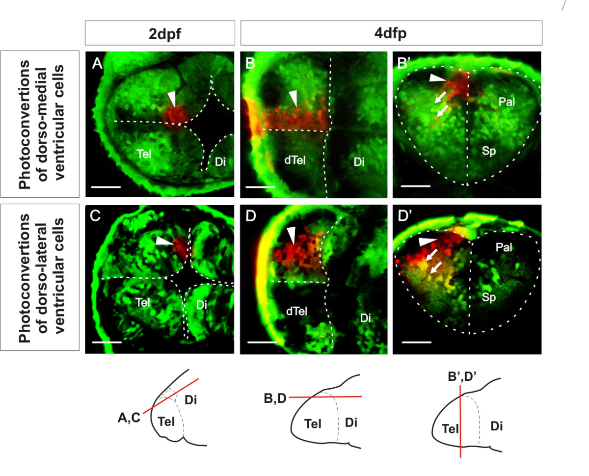
Fig. 6 Ventricular zone at dorsal rim of the AIS expands along the AP axis from 2 dpf to 4 dpf. A. Single confocal z-level in the horizontal plane shows ventricular cells photoconverted (arrowhead) in a medial location of the dorsal rim of the AIS at 2 dpf. B. By 4 dpf, a similar horizontal plane close to the upper (now ventricular) surface of the telencephalon shows that these cells have generated an anteroposteriorly expanded column of cells. B′. A transverse plane through these cells shows that most photoconverted cells are ventricular zone cells that remain close to the sagittal plane of the telencephalic lobe. Arrows indicate radial migration of differentiated neurons. C. Single confocal z-level in the horizontal plane shows ventricular cells photoconverted (arrowhead) in a lateral location of the dorsal rim of the AIS at 2 dpf. D. By 4 dpf, a similar horizontal section close to the upper (now ventricular) surface of the telencephalon shows that these cells have generated an anteroposteriorly expanded column of cells. D′. A transverse section through these cells shows that many of these cells are ventricular zone cells contacting the upper ventricular surface of the telencephalic lobe (arrowhead) and projecting radially (arrows) to the lateral pial surface. Schematics show levels of confocal optical sectioning. Scale bars 50 μm. Di: diencephalon; dTel: upper surface of telencephalon; Pal: pallium; Sp: subpallium; Tel: telencephalon.