
|
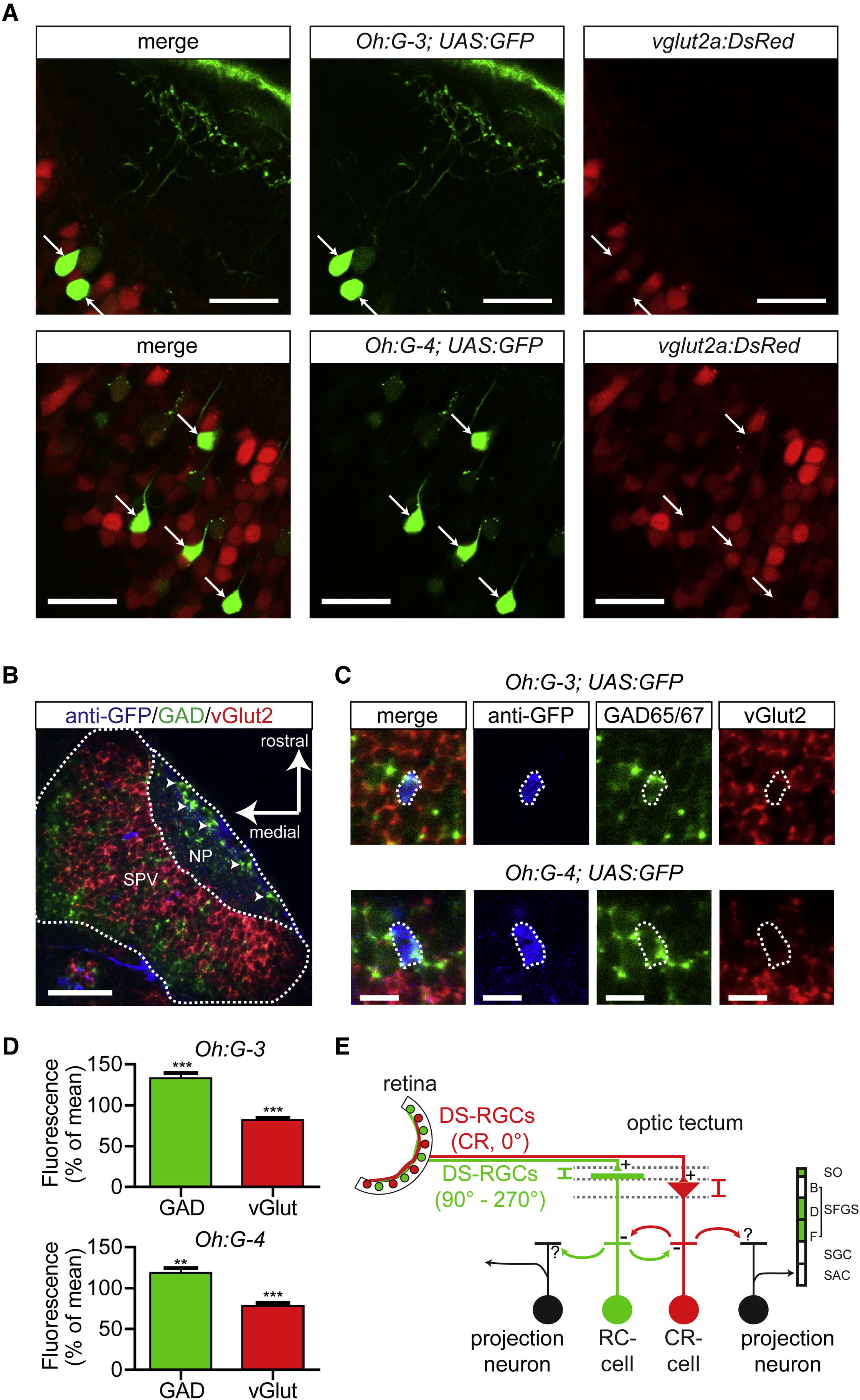
Fig. 7 Neurotransmitter Phenotype of Direction-Selective Neurons in Specific Gal4 Lines(A) Tg(Oh:G-3;UAS:GFP) fish (top row) and Tg(Oh:G-4;UAS:GFP) fish (bottom row) were crossed with Tg(vglut2a:DsRed) fish (confocal images). In both Tg(Oh:G-3) and Tg(Oh:G-4) fish, brightly labeled GFP-positive cells (arrows) were negative for DsRed. Scale bar represents 20 µm.(B) Confocal image of a whole-mount in situ hybridization of a Tg(Oh:G-3;UAS:GFP) larva. NP, neuropil; SPV, periventricular cell body layer. Arrowheads point to GABAergic SINs. Scale bar represents 50 µm.(C) Details of a whole-mount in situ hybridization of Tg(Oh:G-3;UAS:GFP) and Tg(Oh:G-4;UAS:GFP). Scale bar represents 10 µm. Note that anti-GFP-labeled cells (blue) are visible in the green (gad65/67) channel but not in the red (vglut2) channel.(D) In confocal images from the whole-mount in situ hybridization of Tg(Oh:G-3;UAS:GFP) and Tg(Oh:G-4;UAS:GFP), the mean fluorescence intensity within the region of GFP-positive somata in the GAD65/67 channel (green) and vGlut2 channel (red) was measured and normalized to the respective mean fluorescence intensity outside the soma (after background subtraction). In both lines, green fluorescence was significantly higher within GFP-positive somatic regions than outside. Red fluorescence was significantly lower within these GFP-positive somatic regions (Tg(Oh:G-3;UAS:GFP): n = 42 somata; Tg(Oh:G-4;UAS:GFP): n = 40 somata). Error bars indicate mean ± SEM.(E) Schematic drawing representing a possible DS circuit motif in OT. DS type 1 and type 2 cells receive DS excitatory input from layer-specific DS-RGC axons and may provide reciprocal inhibition onto each other and onto other, unidentified tectal neurons.