
|
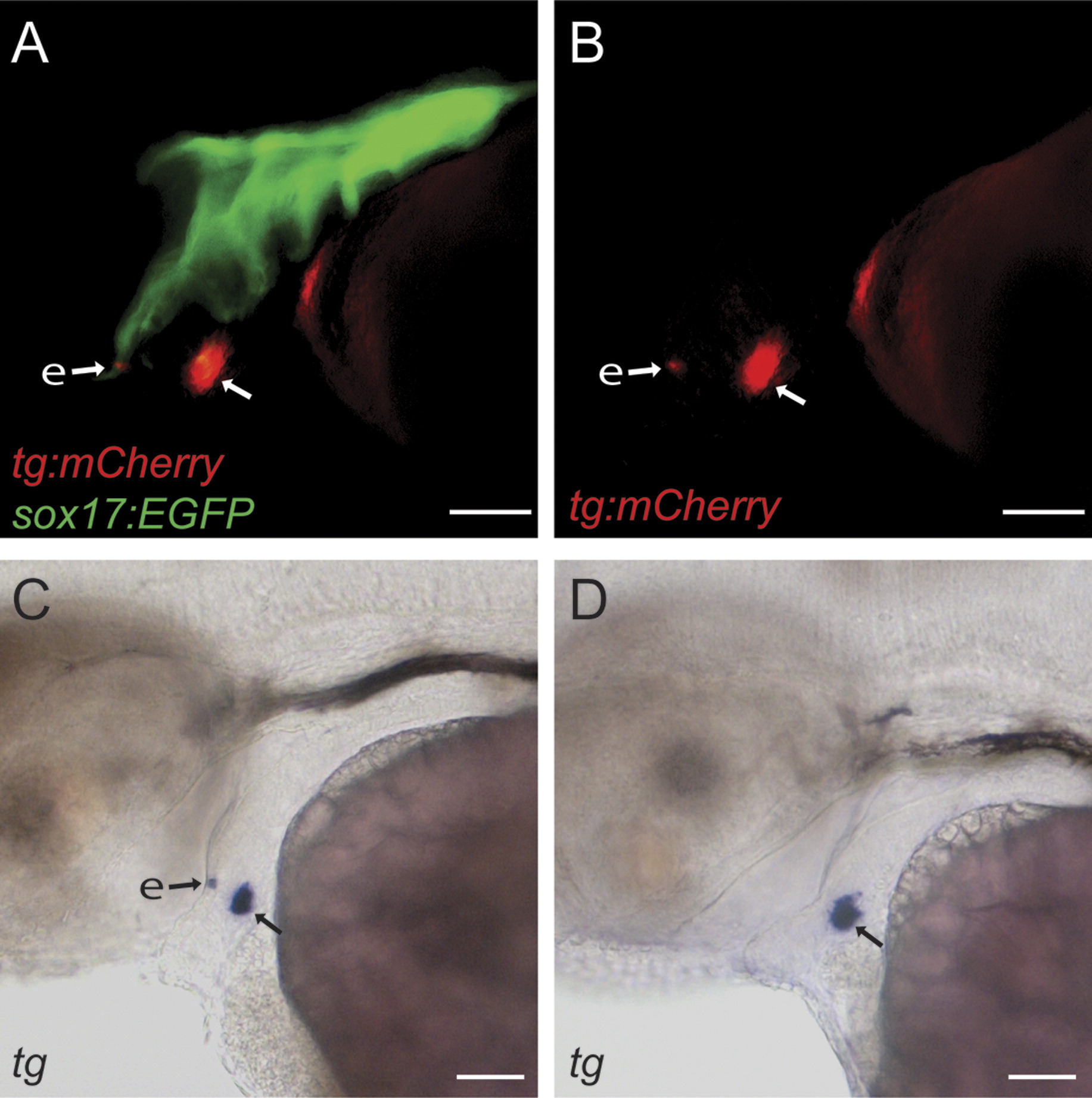
Fig. S1 Detection of ectopic thyroid cells by epifluorescence microscopy in live (tg:mCherry;sox17:EGFP) embryos. Panels A and B show lateral views of a 60 hpf embryo presenting a small group of ectopic fluorescent thyroid cells (e) located within the EGFP-positive pharyngeal epithelium. The main mass of thyroid cells (arrow) showed normal relocation from the pharynx into the subpharyngeal mesenchyme. Panels C and D show results from whole mount in situ hybridization performed with embryos in which ectopic fluorescence was detectable or absent during live imaging. Note that tg mRNA expression could be readily detected within the pharyngeal epithelium of those embryos that displayed an ectopic reporter signal during live imaging (see panel C). In contrast, no ectopic tg mRNA expression was detected in embryos that did not show ectopic ectopic fluorescence during live imaging (see panel D). Scale bar: 50 μM.
Reprinted from Developmental Biology, 372(2), Opitz, R., Maquet, E., Huisken, J., Antonica, F., Trubiroha, A., Pottier, G., Janssens, V., and Costagliola, S., Transgenic zebrafish illuminate the dynamics of thyroid morphogenesis and its relationship to cardiovascular development, 203-216, Copyright (2012) with permission from Elsevier. Full text @ Dev. Biol.