
|
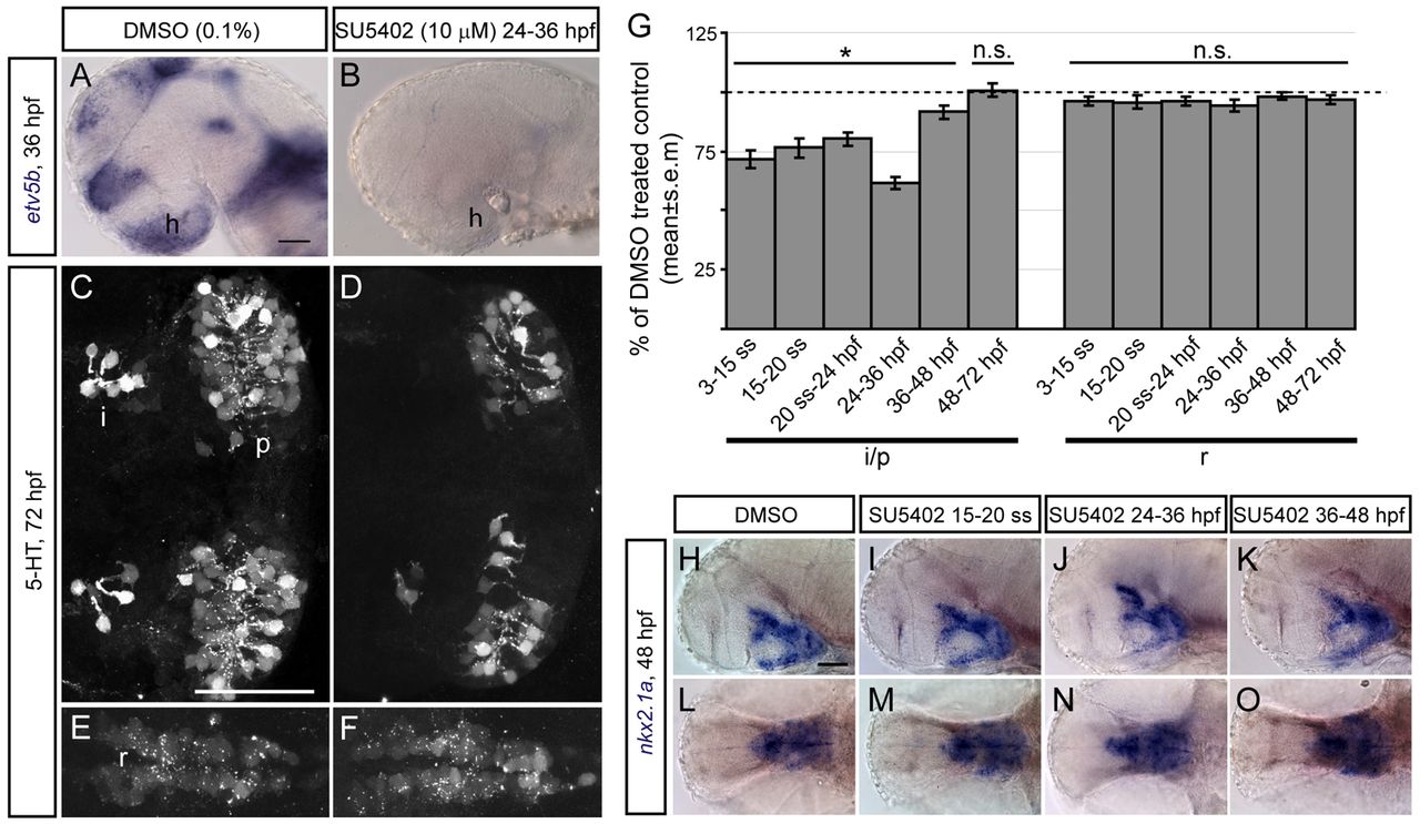
Fig. 5 Development of hypothalamic 5-HT neurons depends on Fgf signalling. (A,B) Micrograph showing DMSO control and SU5402-exposed zebrafish embryos processed for etv5b ISH. Following SU5402 treatment, no etv5b transcripts were detected in the hypothalamus (h). Lateral view, anterior left. (C-F) Confocal maximum intensity projections showing control and SU5402-exposed embryos processed for 5-HT immunohistochemistry. The intermediate/posterior (i./p.) clusters of the hypothalamus (C,D) and the 5-HT population of the anterior raphe (r.) (E,F) are indicated. Ventral views, anterior left. (G) The number of 5-HT cells after SU5402 exposure during the indicated time intervals in clusters i./p. and r. at 72 hpf expressed as percentage of control (dashed line at 100%). *Pd0.05; n.s., not significant. (H-O) nkx2.1a expression in DMSO controls and SU5402-exposed embryos. Lateral (H-K) and ventral (L-O) views, anterior left. Scale bars: 50 μm.