
|
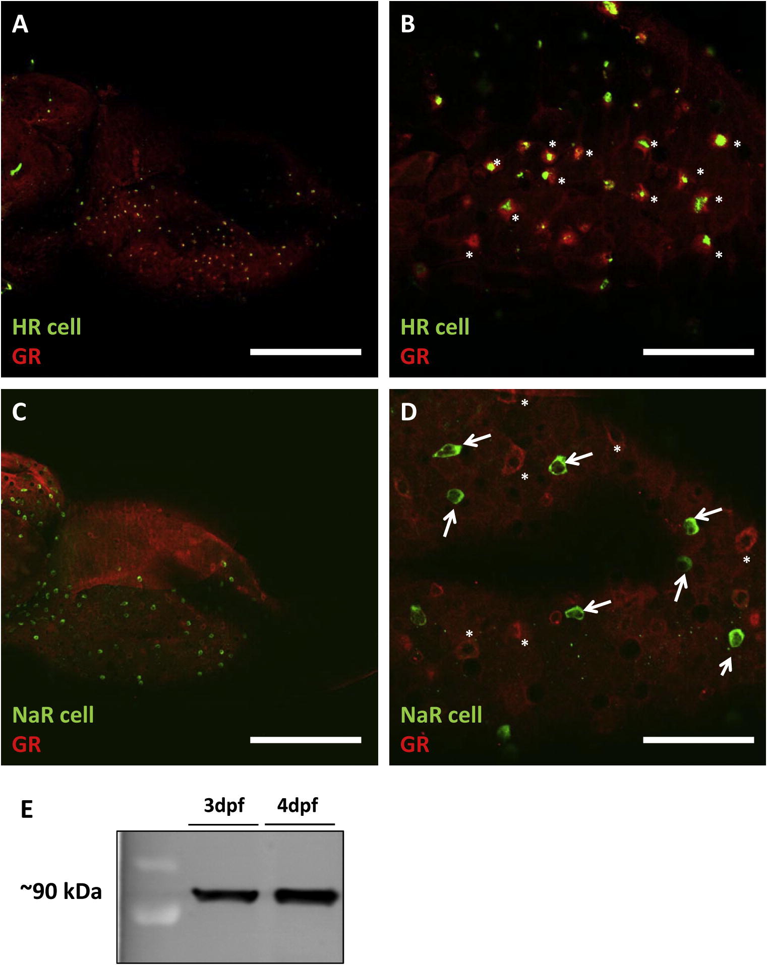
Fig. 6 Localization of GR on yolk sack ionocytes in larval zebrafish. 4 dpf zebrafish larvae were stained with conA (green, a vital dye for HR cells) and GR (red, denoted with asterisks). The majority of GR-positive cells were also stained with conA, suggesting that HR-cells are enriched with GR (A and B). On the other hand, no obvious expression of GR (red, denoted with asterisks) was observed in NaR cells (green; stained with Na+/K+-ATPase antibody, alpha 5, denoted with arrow; (C and D). This antibody successfully recognized an appropriately sized band (<90 kDa) on western blotting (E). Scale bars: 200 μm for Fig. 6A, 6C and 50 μm for (B and D). (For interpretation of the references to color in this figure legend, the reader is referred to the web version of this article.)
Reprinted from Molecular and Cellular Endocrinology, 364(1-2), Kumai, Y., Nesan, D., Vijayan, M.M., and Perry, S.F., Cortisol regulates Na(+) uptake in zebrafish, Danio rerio, larvae via the glucocorticoid receptor, 113-125, Copyright (2012) with permission from Elsevier. Full text @ Mol. Cell. Endocrinol.