
|
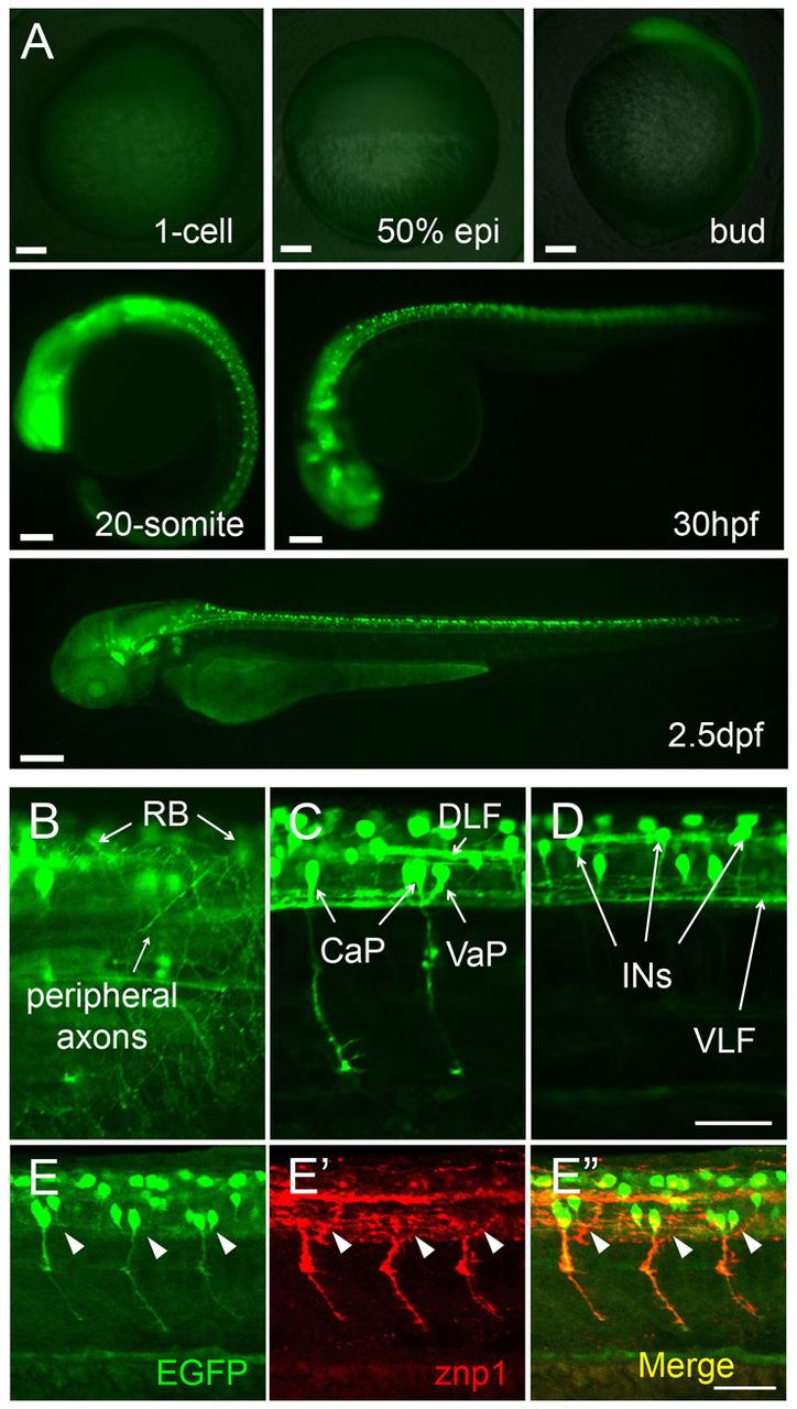
Fig. 1 Neuron-specific expression of GFP transgene in slg+/- zebrafish embryos. (A) GFP expression starts from the bud stage and is restricted to a subset of embryonic neurons. (B-D) GFP expression in RB, CaP, VaP and INs at 32 hpf. (E-E′′) CaP axons labeled by znp1 antibody staining are GFP positive in 26-hpf embryos, whereas MiP axons (arrowheads) are GFP negative. All images are lateral views. Animal pole is to the top for 1-cell and 50% epiboly; dorsal to the right for bud and 20-somite; dorsal to the top and anterior to the left for 30 hpf and 2.5 dpf in A and for B-E′′. CaP, caudal primary motoneurons; DLF, dorsal longitudinal fasciculus; INs, interneuron-like cells; RB, Rohon-Beard neurons; VaP, variable primary motoneurons; VLF, ventral longitudinal fasciculus. Scale bars: 100 μm in A; 50 μm in B-E′′.