
|
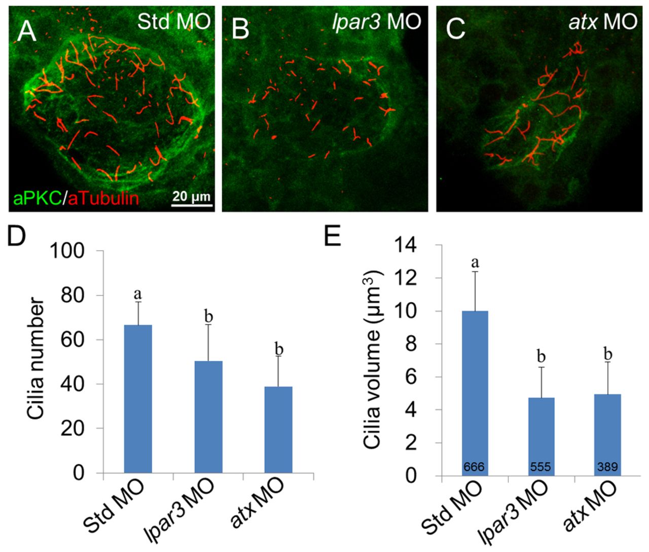
Fig. 5 KV ciliogenesis is disturbed in lpar3 and atx morphants. (A-C) Cilia in KV for the indicated treatments were observed by immunofluorescence using anti-acetylated tubulin antibody (red) and counter-stained with aPKC antibody (green) to outline the KV. Representative photographs of Std MO-injected zebrafish embryos (A), lpar3 (B) and atx (C) morphants are presented (anterior side up). (D,E) Ten embryos were used for each treatment (three independent experiments) to measure cilia number (D) and volume (E). The number of embryos examined is presented in each column. Error bars represent s.e.m. a, b, groups denoted with different lettering refer to statistical significance (P<0.05).