
Image
|
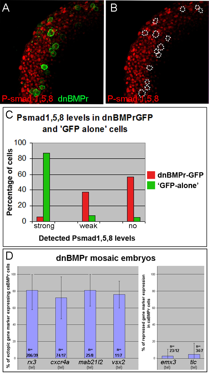
Figure Caption
Fig. s5 Majority of dnBMPr transgenic cells have no or low levels of P-Smad1,5,8 (A,B) Animal view of the ventral side of a mid gastrula dnBMPr mosaic embryo stained for Psmad1,5,8. (B) Dotted circles mark the location of dnBMPr expressing cells (green in A). (C) Distribution of P-smad1,5,8 signal intensities in dnBMPr (n=128 cells) and GFP-alone (n=148 cells) expressing cells. Statistical analysis of induced and repressed gene expression in dnBMPr cells, respectively. n=number of cells/ number of embryos. (tel, telencephalon)
Acknowledgments
This image is the copyrighted work of the attributed author or publisher, and
ZFIN has permission only to display this image to its users.
Additional permissions should be obtained from the applicable author or publisher of the image.
Reprinted from Developmental Cell, 23(4), Bielen, H., and Houart, C., BMP Signaling Protects Telencephalic Fate by Repressing Eye Identity and Its Cxcr4-Dependent Morphogenesis, 812-822, Copyright (2012) with permission from Elsevier. Full text @ Dev. Cell