
|
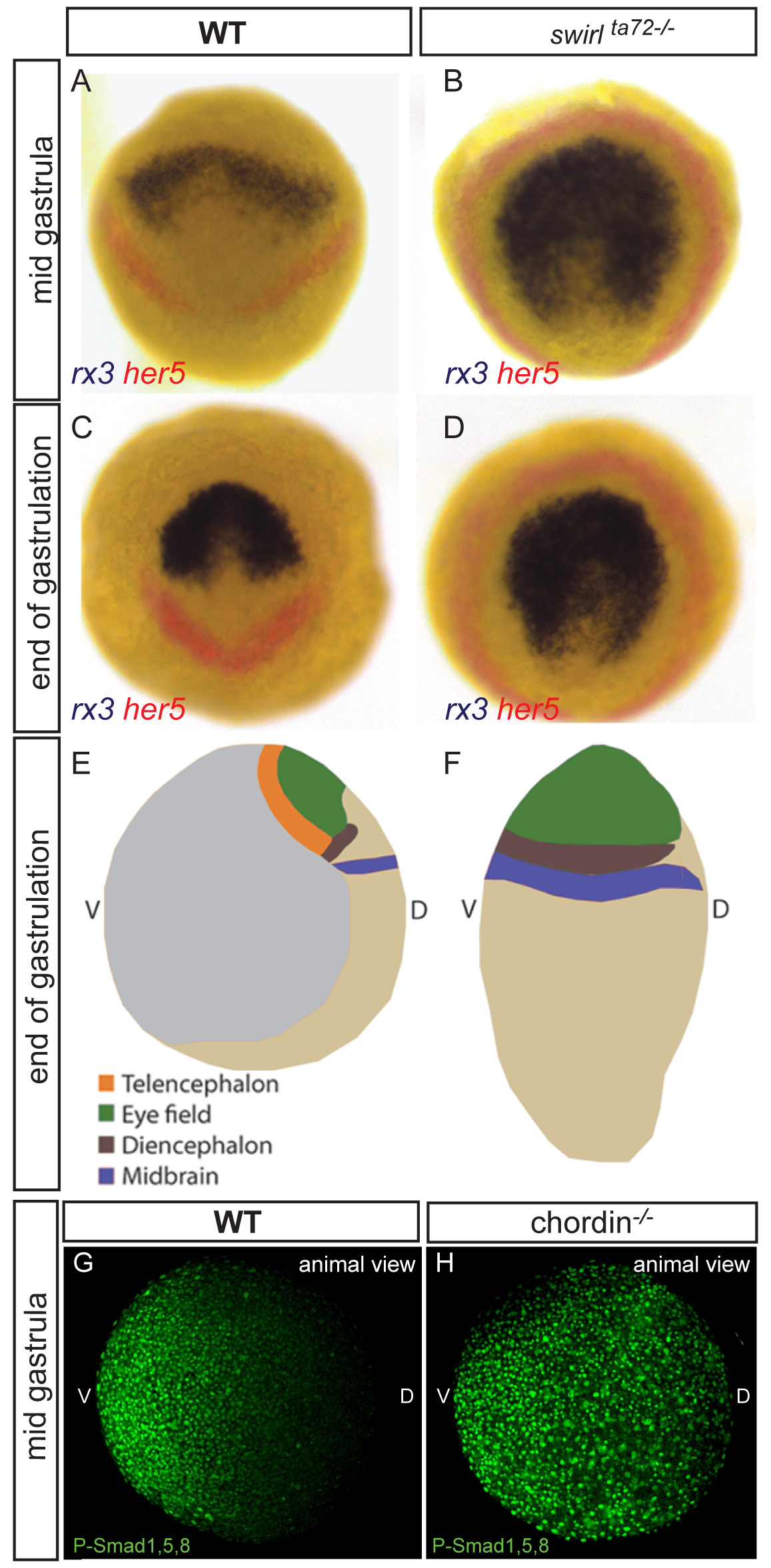
Fig. s1 Double in situ of early eye-field (rx3) and midbrain (her5) markers in WT (A,C) and swr72-/- (B,D) embryos at 75% epiboly (A,B) and bud stage (C,D). Dorsal view (A,C) and animal view (B,D). (E,F) Schematic overview of changes of expression pattern in WT and swr72-/- at bud stage. The telencephalon at the margin of the neural plate is the only dorsal, neural region which is not expanded but absent in swr72-/-. Lateral view, dorsal to the right, anterior to the top. (G,H) Detection of P-smad1,5,8 in WT (G) and chordin-/- (H) embryos, animal pole view, shield to the right. Figures are z-projections of confocal sections.
Reprinted from Developmental Cell, 23(4), Bielen, H., and Houart, C., BMP Signaling Protects Telencephalic Fate by Repressing Eye Identity and Its Cxcr4-Dependent Morphogenesis, 812-822, Copyright (2012) with permission from Elsevier. Full text @ Dev. Cell