
|
Fig. 6
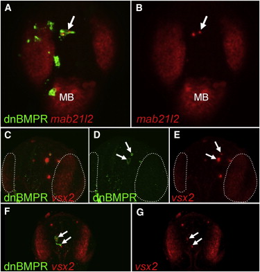
Ectopic Expression of Retinal Markers in BMP-Depleted Telencephalic Cells at 24 hpf (A–G) Embryos were injected with HSP70:dnBMPr plasmid at one- to two-cell stage followed by heat shock at oblong/early blastula stage. The arrows indicate dnBMPr-positive cells in the telencephalon at 24 hr, which ectopically express retinal markers mab21l2 (A and B) and vsx2 (C–G). The number of transiently transgenic dnBMPr cells is significantly lower at 24 hpf than at earlier stages, and the remaining transgenic cells frequently undergo apoptosis (arrows in D). (C–E) White dotted lines mark the vsx2-positive retina. All views are dorsal, anterior at the top. All figures are z-projections of confocal sections. MB, midbrain.
Reprinted from Developmental Cell, 23(4), Bielen, H., and Houart, C., BMP Signaling Protects Telencephalic Fate by Repressing Eye Identity and Its Cxcr4-Dependent Morphogenesis, 812-822, Copyright (2012) with permission from Elsevier. Full text @ Dev. Cell