
|
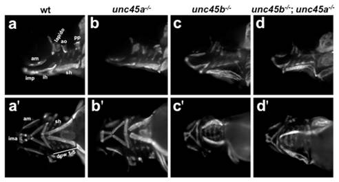
Fig. 5 Myosin localization in the craniofacial muscles of 4 dpf embryos.
Wild type siblings (a,a′); unc45a-/- (b,b′); unc45b-/- (c,c′); unc45b-/-; unc45a-/- (d,d′) mutants. Lateral (a-d) and ventral (a′–d′) views, head to the left. Expression is unaltered between wild type siblings (a,a′) and unc45a-/- (b,b′), unc45b-/- (c,c′) or unc45b-/-; unc45a-/- (d,d′) mutants. The sternohyoideus is displaced in unc45b-/- (c,c′) and unc45b-/-; unc45a-/- (d,d′) embryos. Primes denote alternative views of the same embryo. ao, adductor operculi; am, adductor mandibulae; do, dilator operculi; dpw 1–5, dorsal pharyngeal wall; hh, hyohyoideus; ih, interhyoideus; ima, intermandibularis anterior; imp, intermandibularis posterior; lap, levator arcus palatini; pp, pterygoid process; sh, sternohyoideus.