
|
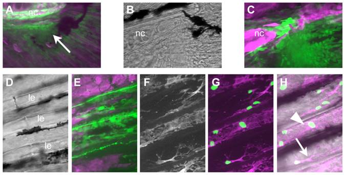
Fig. 8 The scleroblasts of the caudal fin are NC derived.
A) At 8 dpf, NC-derived cells (GFP+; arrow) can be seen clustered around the tip of the notochord (nc). B, C) By 16 dpf, there are more GFP+ cells; some are located more distally in the fin, although many are still close to the notochord. D-H) At 21 dpf, the caudal fin contains well-formed lepidotrichia (le in D), which are associated with GFP+ cells (E). F-H) To confirm the identity of the cells as osteoblasts, fish carrying the nucCh reporter were crossed with RUNX2:egfp transgenics, in which the osteoblasts are GFP+. The osteoblasts have nucCh+ nuclei, indicating they are NC-derived (G), and they are located both within (arrowhead) and immediately outside (arrow) the lepidotrichia (H).