
|
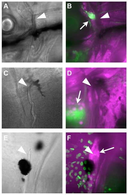
Fig. 7 NC does not contribute to the cleithrum.
A-D) At 10 dpf (A, B) and 16 dpf (C, D), Z-stack projections of confocal sections through the area surrounding the dorsal end of the cleithrum (arrowheads) reveal no GFP+ cells. The NC-derived glia of the lateral line ganglia are clearly visible in the same fields of view (arrows). E, F) To localize the osteoblasts, fish carrying the nucCh reporter were crossed with RUNX2:egfp transgenics, in which the osteoblasts are GFP+. The dorsal tip of the cleithrum (arrowhead in E, F) is surrounded by osteoblasts (arrow in F), which do not have nucCh+ nuclei, indicating they are not NC-derived.