
|
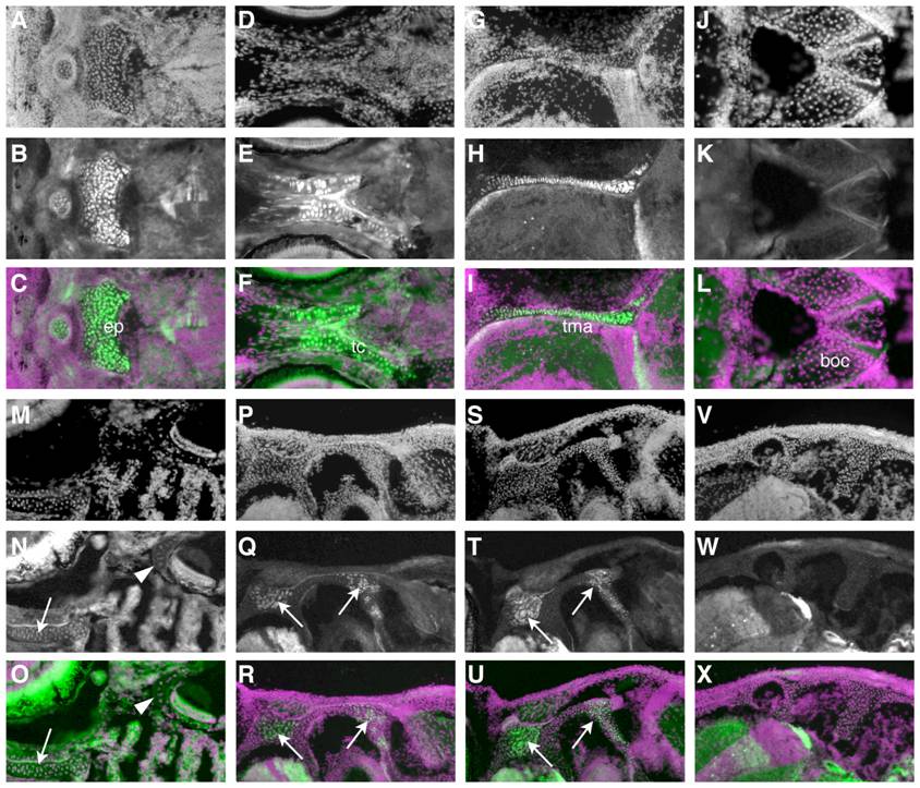
Fig. 4 The chondrocranium is of mixed origin.
A-R) Immunohistochemistry for GFP shows composition of cartilages with cellular resolution. In each set of three images, the first shows the DAPI counterstain (A, D, G, etc.), and the second (B, E, H, etc.) the GFP immunoreactivity. The third image in each group, the overlays, are pseudocolored with green representing GFP immunoreactivity and magenta the DAPI counterstain. The most anterior cartilages in the base of the skull, such as the ethmoid plate (A-C), trabeculae cranii (D-F), and taeniae marginalis anterior (G-I) are NC-derived. More posterior cartilages, like the basioccipital (J-L), contain no NC. M-O) A horizontal section at 14 dpf illustrates a more anterior NC-derived cartilage (arrow), the trabeculae cranii, and more posterior negative cartilage around the ear (arrowhead). P-X) Successive sections through a single fish at 44 dpf show that cartilage at intermediate locations, such as around the ear, is composed of a mix of NC (arrows) and non-NC cells in more ventral sections (P-R), and shows no NC-derived cells more dorsally (V-X).