
|
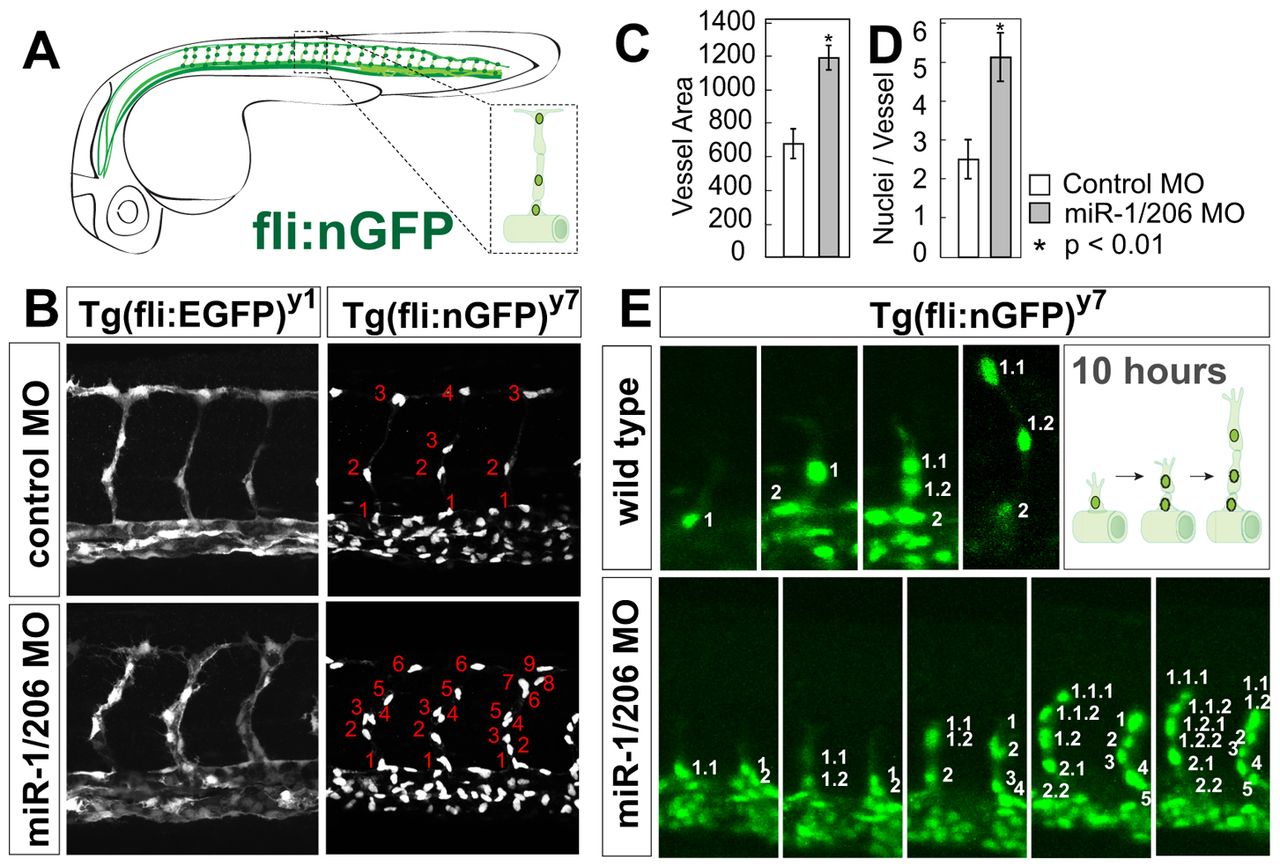
Fig. 1 miR-1 and miR-206 regulate developmental angiogenesis. (A) Schematic representation of the zebrafish vasculature. The intersegmental vessels (ISVs) form by sprouting endothelial cells from the dorsal aorta and are typically composed by three cells: the tip, the stalk, and the basal cell that connects to the aorta (inset). (B) ISVs labeled with cytoplasmic GFP [Tg(fli-EGFP)y1] or nuclear GFP [Tg(fli-nGFP)y7] after injection of control morpholino (MO) or miR-1/206 MO at the one-cell stage. (C,D) Quantification of the cross-sectional area (C) and average number of nuclei per ISV (D) in control MO-injected and miR-1/206 MO-injected embryos at 30 hpf. Note the increase in ISV size (cross-sectional area) and number of endothelial cells in embryos injected with miR-1/206 MO compared with control MO-injected embryos. Mean ± s.d.; Student′s t-test. (E) Individual frames of a time-lapse confocal analysis of the vascular phenotype in wild-type and miR-1/206 MO-injected embryos between 18 and 28 hpf. Endothelial cells migrating into the vessel are numbered (1, 2, 3, etc.) and their daughter cells are labeled (1.1, 1.2, 1.2.1, etc.). The wild-type ISV shows a highly stereotyped pattern, in which a single endothelial cell migrates to the trunk midline and divides, leaving one cell stationary while the tip cell continues to migrate dorsally. Endothelial cells in miR-1/206 MO-injected embryos show elevated proliferation within the ISV (left vessel), as well as an increase in migration from the aorta (right vessel).