
|
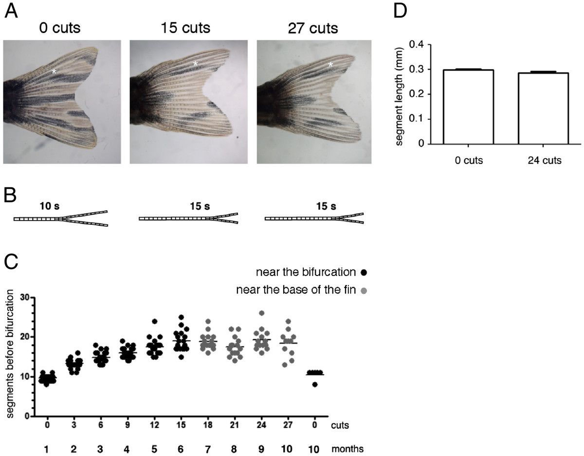
Fig. 1 The bifurcation position is distalized with repeated amputations. (A) The same caudal fin before amputation and after 15 and 27 amputations. (B) Schematic representation of the bifurcation distalization with the repeated amputations. (C) Number of segments formed in the 3rd dorsal ray between the base of the fin and the bifurcation after consecutive amputations. The caudal fin was subjected to three amputations every month and this was repeated 10 times. During the first 15 amputations, the third consecutive amputation (the last before allowing the fin to completely regenerate) was done three segments below the most proximal bony ray bifurcation (near the bifurcation). In the next 12 cuts, the third consecutive amputation was done 4 segments distally from the base of the fin (near the base of the fin). (D) 3rd dorsal ray segment length before any amputation and after 24 amputations. Asterisk marks the bifurcation position. The fins were allowed to regenerate for 4 weeks between each round of three consecutive amputations.